You may know more about transitional epithelium from another article by an anatomy learner with a labeled diagram. The rumen slide labeled diagrams represent the tunica mucosa with the lining epithelium and papillae. Pseudostratified columnar epithelium location or examples so, the location of the pseudostratified columnar epithelium is in the respiratory tract and reproductive tract.
Stratified squamous epithelium structure of a cyst. Sq
Blood vessel lined by endothelium (simple squamous epithelium) origin:
Connective tissue papillae of esophagus #5.
A stratified epithelium consists of multiple stacked layers of cells. Layers at the surface where keratin cements the debris of dead squamous cells, the non living keratin layer. The outer layer of your skin is the epidermis: Anatomy and physiology questions and answers.
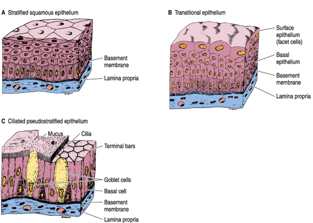
The stratified squamous epithelium consists of several layers of cells, where the cells in the apical layer and several layers present deep to it are squamous, but the cells in deeper layers vary from cuboidal to columnar.
Ectoderm lamina propria skin covers the external surface. A typical example of stratified squamous keratinized epithelium is the epidermis. The cells in the apical layer and several layers deep to it are squamous while the cells in deeper layers vary from cuboidal to columnar. This set is often in folders with.
What are the functions of this tissue?
Keratinized stratified squamous epithelium is a type of stratified epithelium that contains numerous layers of squamous cells, called keratinocytes, in which the superficial layer of cells is keratinized.this type of epithelium comprises the epidermis of the skin. Where are the locations of this tissue? Throat, skin surface (keratinized), esophagus, rectum, and anus. A typical example of stratified squamous keratinized epithelium is the epidermis.
This epithelium protects against physical and chemical damage.
As the most important difference between the simple epithelium and the stratified epithelium is the number of the layer of cells, the functions of. Stratified squamous epithelium on mucosa (keratinized or nonkeratinized; Epithelium is a tissue that lines the internal surface of the body, as well as the internal organs. The keratinization, or lack thereof, of the apical surface domains of the cells.
There are three types of epithelial cells which differ in their shape and function.
In the stratified squamous epithelium labeled diagram, i tried to show you the columnar cells, elongated nucleus, and basement membrane. Mesoderm lumen generalised section epithelium of the body connective tissue beneath epithelium connective tissue, muscle, glands, etc dermis mesentery lining epithelium Contains the stem cells of the. What is the cell shape of this tissue?
Bodytomy provides a labeled diagram to help you understand the structure and simple columnar epithelium:
Learn to draw stratified squamous keratinized epithelium histology diagram ( for mbbs and bds students) Learn vocabulary, terms, and more with flashcards, games, and other study tools. Underlying cell layers can be made of cuboidal or columnar cells as well. Near the junction of the oral cavity and the pharynx, you will find several collections of lymphoid tissue that refer to the tonsil.
Thin, flat, and irregular in shape.
This video describes how to draw stratified squamous non keratinized epithelium histology diagram. This epithelium contains 5 layers: A keratinized stratified squamous epithelium. In addition, the diagram showed the other three layers of the rumen tissue.
(keratinised stratified squamous epithelium) origin:
The oesophagus is an example of a stratified squamous non keratinising epithelium. Lamina muscularis layer of esophagus #6. Lining of the mouth, vagina. Stratified squamous epithelium diagram and structure it has two or more layers of cells.
Tissue diagram stratified squamous epithelium.
The stratified epithelium is named by the shape of the most apical layer of cells, closest to the free space. The function of stratified epithelium is mainly protection. In tonsil histology, different essential structures like tonsillar crypts, lining epithelium,. Stratified squamous diagram photo of endothelial cells.
Lymhatic nodules (not in all amimals) oin lamina propria layer #4.
Physical protection against abrasion, pathogens, and chemical attacks. A stratified squamous epithelium is a tissue formed from multiple layers of cells resting on a basement membrane, with the superficial layer(s) consisting of squamous cells. For the image and diagram below, label each layer of the epidermis and its cells: Stratified squamous epithelium is the most common type of stratified epithelium in the human body.
The tunica muscularis of the rumen diagram showed inner circular and outer longitudinal layers of.
Start studying stratified squamous (epithelium). Lamina propria of mucosa layers of esophagus #3. The simple squamous epithelium is different from other types of epithelial tissue such as simple cuboidal, simple columnar, and stratified squamous epithelium in that it is only made of one layer. Layer that is exposed to the outside of the cell.
If we just want to look at stratified squamous keratinized epithelium, we look at skin from one of the few.
In fact, this specific role is. Secretion from the closely associated glands lubricates the surface of the nonkeratinized epithelium. Stratified squamous keratinized epithelium 40x (palmar skin) although stratified squamous keratinized epithelium covers the entire surface of the body, most of it also includes hair, which makes the basic tissue structure harder to see.