Researchers do know that it produces and. Erik maronde, a survey of molecular details in the human pineal gland in the light of phylogeny, structure, function and chronobiological diseases, journal of pineal. [article in russian] author a a maksimovich.
Video/Image Link Pineal Gland
Basically, the structure of the pineal gland is 0.3 inches long and 0.1 grams of weight.
These fibers contain substances including:
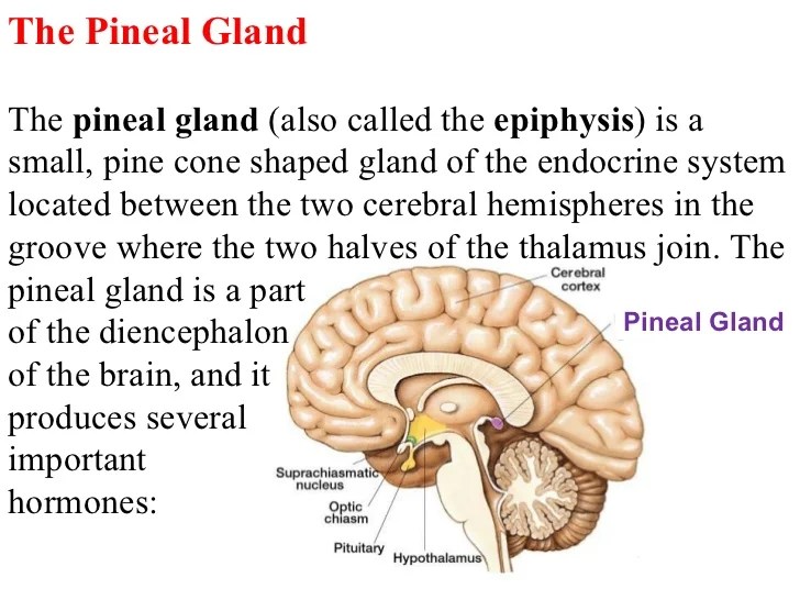
It is a neuroendocrine gland that secretes the hormone melatonin and several other polypeptide hormones that have a regulatory function on other endocrine glands. The pineal gland is about 0.3 inches long and weighs 0.1 grams. The pineal gland is a midline structure, located between the two cerebral hemispheres. The adrenergic nerves entering the pineal gland regulate its functions.
Since the gland is present at the posterior area of the cranial fossa in the brain, it is also known as the epiphysis cerebri.
Structure pineal gland function the primary function of the pineal gland is to produce melatonin, which has various functions in the central nervous system, the most important of which is to help modulate sleep patterns. The pineal stalk carries nerve fibers to the gland. Its function isn’t fully understood. Melatonin, the main pineal secreting hormone, has been extensively studied in primary and secondary headache disorders.
The pineal gland is a relevant structure and linked to different situations.
The pineal gland plays an important role in biological rhythms, circadian and circannual variations, which are key aspects in several headache disorders. The pineal gland the function of this small organ near the center of the mammalian brain has long been a mystery. The pineal gland is composed of pinealocytes and supporting cells that resemble the astrocytes present in the brain. Sleep patterns are considered to be modulated by this hormone as its production is stimulated by the absence of light.
The pineal gland has a very unique cellular structure.
As it is one of the endocrine glands it secretes its product, the hormone called melatonin, directly into the blood. The pineal gland is composed of cells called pinealocytes and cells of the nervous system called glial cells. Department of anatomy, university of texas health science center at san antonio, san antonio, texas 78284. The pineal gland is an unpaired epithalamic structure, originating as an evagination of the roof of the third ventricle and from the same embryological tissue from
It is attached by a stalk to the posterior wall of third ventricle.
The pineal gland was described as the “seat of the soul” by renee descartes and it is located in the center of the brain. The pineal gland is an endocrine gland present at the geometric center of the brain that is essential in the circadian cycle of sleep and wakefulness of the body. Altered melatonin secretion occurs in many headache syndromes. Recent studies indicate that it is a biological clock that regulates the activity of the sex glands by richard j.
The pineal gland is an endocrine gland located in the epithalamus, near the center of the human brain.
Being part of both the nervous system and the endocrine system, its basic functioning is the emission of various hormones that will alter different brain and other body systems. Its main cell type are pinealocytes which are unique cells to the pineal gland. Neurotransmitters are chemical messengers released by nerve cells that influence pinealocytes. The pineal gland is made up of pinealocytes and supporting cells that manage the functions of astrocytes and give commands to the brain.
[structure and function of the pineal gland in the vertebrates] [structure and function of the pineal gland in the vertebrates] zh evol biokhim fiziol.
Wurtman and julius axelrod buried nearly in the center of the brain in any mammal is a small Pineal gland structure in mammals and humans, pinacocytes and interstitial cells both produce hormones within the pineal gland.


