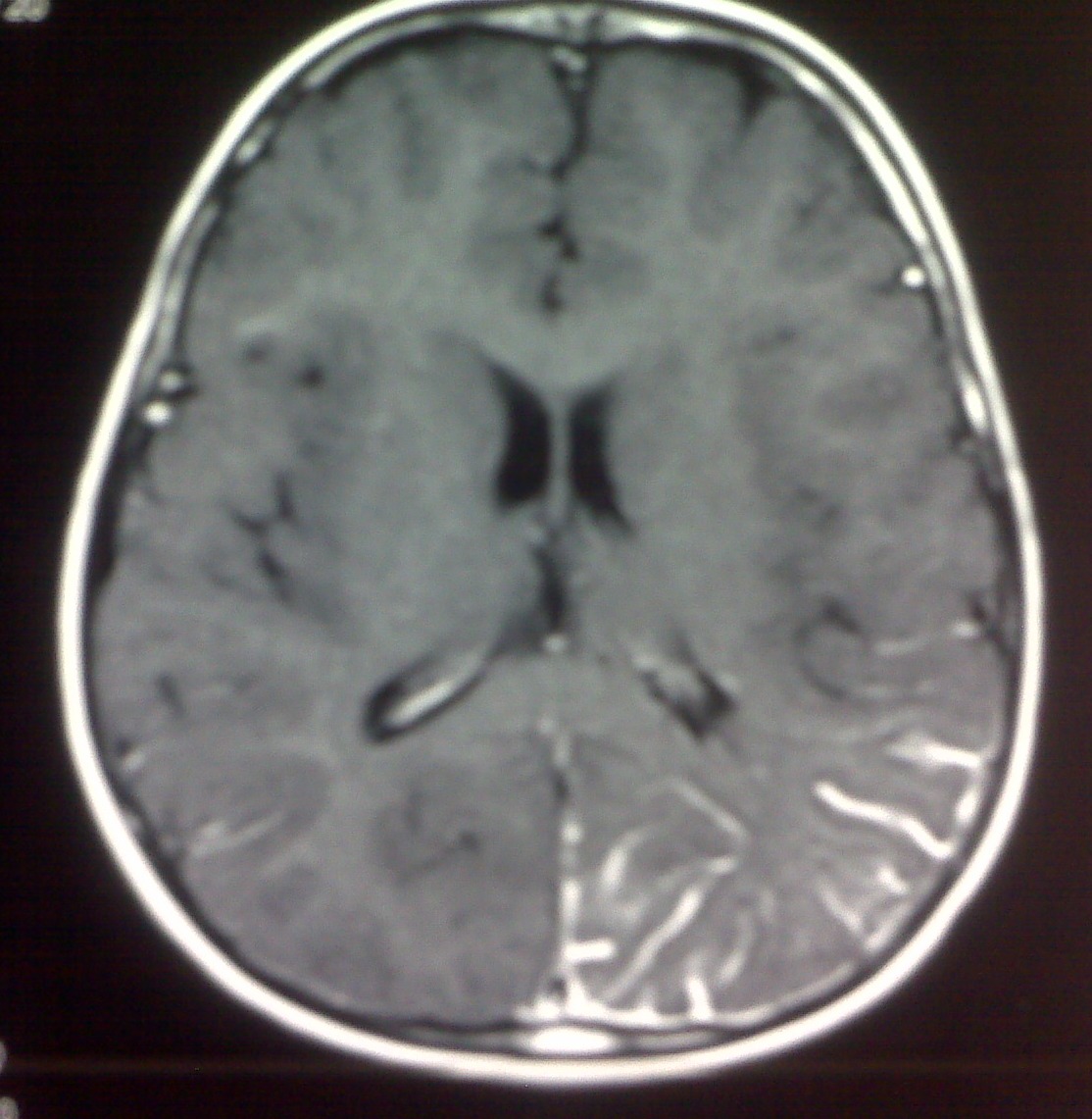
Summarising their recent work, presented at the society for interventional radiology 2019 conference ( sir; The affected hemisphere's image is smaller, the overlying cap widened, and both more radioactive than the uninvolved side. 1 at birth or during childhood, the capillary malformations.
KlippelTrénaunayWeber syndrome Radiology Case
The exact pathophysiology and genetic etiology of the disorder are unknown.
Neurocutaneous syndromes are a diverse group of disorders that involve both skin and nervous systems.
There is a thickening of right calvarial thickening along with hyperpneumatisation of right frontal sinus. Based on the extent of leptomeningeal. “port wine” vascular nevus flammeus in the trigeminal nerve distribution; Occasionally the substantia nigra can also be involved 5.
Evolution of radiological findings in the adulthood variant of sws with isolated leptomeningeal angiomatosis has never been reported to our knowledge.
In cerebral calcification regions, patients frequently displayed. It has been postulated that the presence of facial and pial angiomas suggests the persistence of primordial sinusoidal vascular channels. 35 year male presented with h/o frequent fall , seizures & abnormal cognitive state. Intracranial angiomatosis is confined to the pia mater.
The prevalence of ktws is 1 :
It is imperative that both the radiologist and surgeon be aware of this entity, as. Prenatal diagnosis using ultrasound has been reported. Clinical and mri data were analyzed. Possible associated cleral and choroidal.
Occasionally the substantia nigra can also be involved 5.
Nellhaus g, haberland c, hill bj. For the purpose of describing the imaging findings and elucidating the role of medical imaging in the diagnosis and assessmen. On ct, extensive gyral and subcortical calcification is seen at right cerebral parenchyma along with right cerebral atrophy. It is typically located in the occipital lobe.
We studied 14 consecutive cases with clinical and radiological evaluations [computed tomography (ct) and magnetic resonance imaging (mri)].
Lapras c, dechaume jp, revol m, nicolas a, deruty r.


