Tuberous sclerosis or tuberous sclerosis complex (tsc) is a genetic disorder that is characterised by hamartomas in many organs, but particularly the skin, brain, eye, kidney and heart. Computed tomography (ct) and magnetic resonance (mr) imaging findings were reviewed in 26 patients with tuberous sclerosis. We review the computed tomography (ct) and magnetic resonance (mr) features of the brain lesions in patients with tuberous sclerosis.
Lipoma in a patient with tuberous sclerosis. CT scan
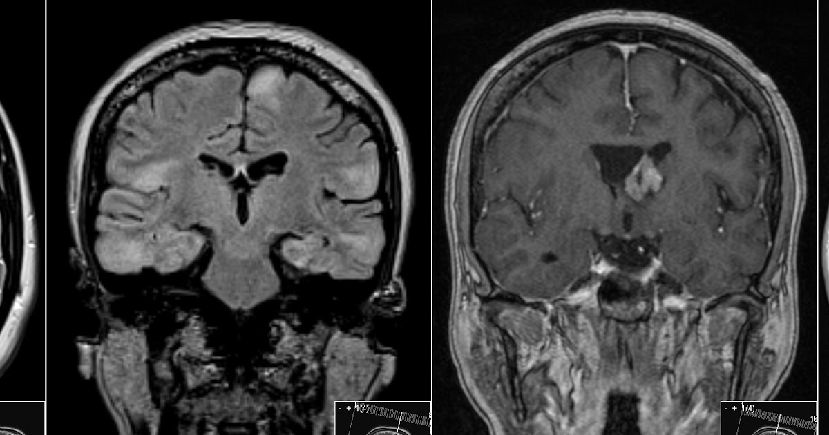
Ct image shows calcified subependymal nodules in right caudothalamic groove (arrow) and posterolateral to right thalamus (arrowhead).
The ct findings in a patient with tuberous sclerosis are described with special emphasis upon the differential diagnosis.
57 rows tuberous sclerosis complex (tsc) is characterized by the growth of benign tumors throughout the body, including in the heart, brain, and kidneys. Mr imaging demonstrates more clearly cortical, and white matter lesions than ct, since mr imaging shows excellent image contrast between various normal structures and high. Frequently, ct can depict extracranial manifestations of tuberous sclerosis. Tuberous sclerosis is a rare genetic disorder that causes noncancerous (benign) tumors ― unexpected overgrowths of normal tissue ― in parts of the body.
Parenchymal hamartomas (cortical tubers) were seen in 23 of 26 patients (88%).
A combination of symptoms may include seizures, intellectual disability, developmental delay, behavioral problems, skin abnormalities, lung disease, and. Advice, updates and vaccine options Us, ct, and mr diagnosis of brain and cardiac lesions pediatr radiol. The presence of multiple bilateral subependymal nodular nonenhancing hyperdense calcified lesions is relatively characteristic of tuberous sclerosis when combined with the appropriate clinical findings.
It usually affects the central nervous system and can result in a combination of symptoms including seizures, impaired intellectual.
Ct clearly demonstrates calcified subependymal nodules. Describe the advantages and disadvantages of the alternative techniques for diagnosis of tuberous sclerosis. The presence of multiple bilateral subependymal nodular nonenhancing hyperdense calcified lesions is relatively characteristic of tuberous sclerosis when combined with the appropriate clinical findings. Tuberous sclerosis complex (tsc) is an autosomal dominant multisystemic disorder characterized by the formation of hamartomas and dysplastic lesions in various organs such as skin, heart, kidneys, brain, and lungs (henske, jozwiak, kingswood, sampson, & thiele, 2016).
Tuberous sclerosis (ts) is an autosomal dominant inherited neurocutaneous syndrome characterized by a variety of hamartomatous lesions in various organs.
The ct features included subependymal nodules in 25 of 26 patients (96%) and calcifications in 23 of 26 (88%). Tuberous sclerosis complex (tsc) is an autosomal dominant disorder characterized by hamartomas in one or many organs, particularly in the skin, central nervous system, retina, and kidneys. Subependymal nodules are seen in nearly all tuberous sclerosis patients and calcify as patient ages. Tuberous sclerosis complex (tsc) is a genetic disorder affecting cellular differentiation, proliferation, and migration early in development, resulting in a variety of hamartomatous lesions that may affect virtually every organ system of the body.
Overall, ct reveals intracranial abnormalities in 85% of patients with tuberous sclerosis.
1a —cns manifestations of tuberous sclerosis. This happens when cells grow out of control and divide more than they should. There are multiple and bilateral small cortical renal lesions of fatty density in keeping with renal angiomyolipomas. Two hyperdense renal cysts are noted with an average attenuation of 45 hu on nonenhanced ct (the cystic nature was confirmed by ultrasound).
Certain symptoms develop before to birth, such as heart tumors (rhabdomyoma).
Ct is superior to mr in demonstrating the presence of subependymal nodules, thereby confirming or establishing the diagnosis of tuberous sclerosis. Ct readily depicts calcified cortical tubers and calcified subependymal nodules; To determine if sclerotic bone lesions evident at body computed tomography (ct) are of value as a diagnostic criterion of tuberous sclerosis complex (tsc) and in the differentiation of tsc with lymphangioleiomyomatosis (lam) from sporadic lam. Other symptoms become more obvious in childhood, such as developmental delay and skin changes.
The ct findings in a patient with tuberous sclerosis are described with special emphasis upon the differential diagnosis.
Ct examination revealed fibrous dysplasia involving the frontal, ethmoid, sphenoid, and vomer bones. Authors c christophe 1 , j bartholome, d blum, a clerckx, b desprechins, d de wolf, a gallez, p. Hamartomas can grow in many parts of the body. Pulmonary involvement has been reported to.