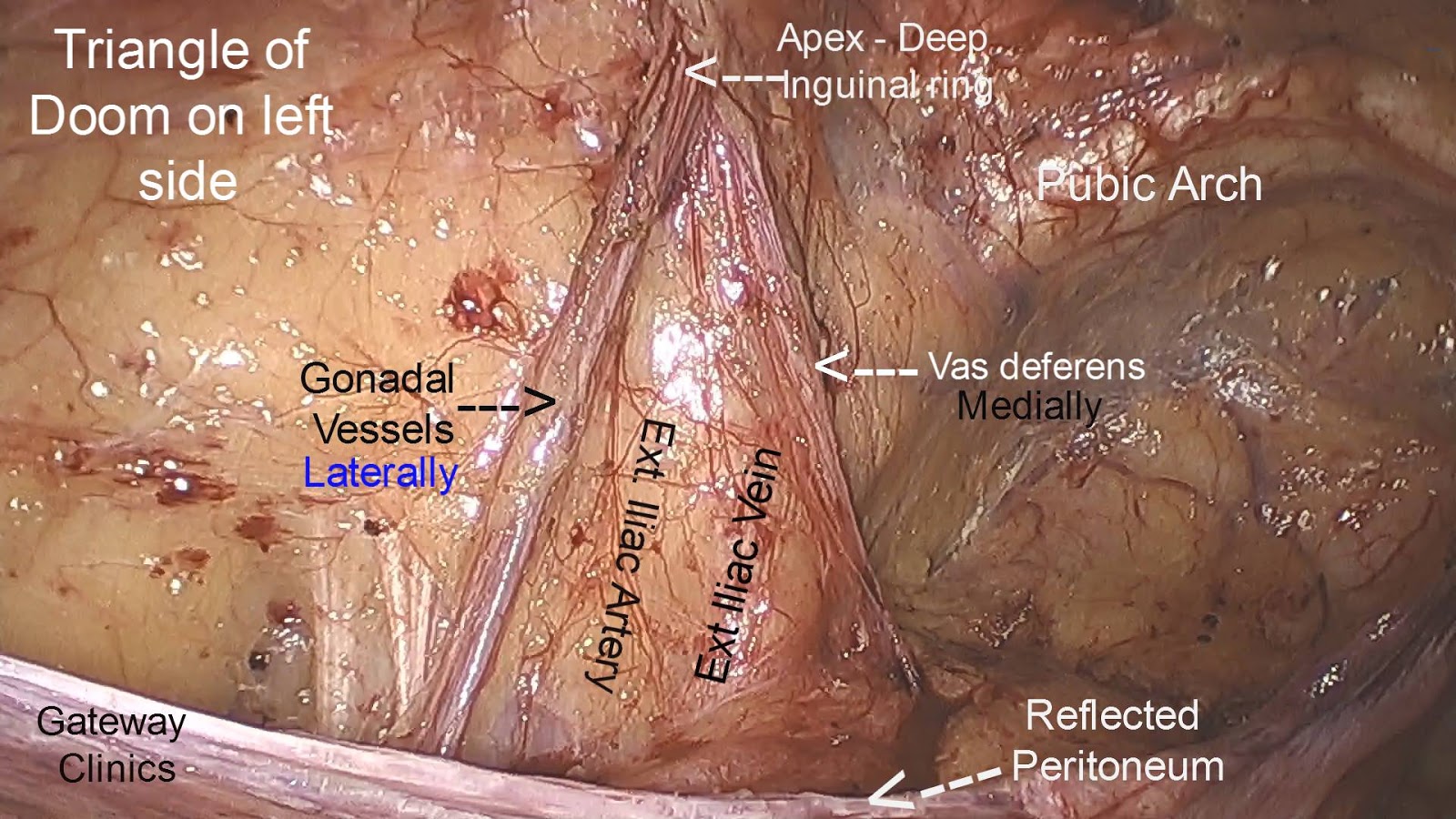
The triangle of doom is bordered medially by the vas deferens and laterally by the vessels of the spermatic cord, the summit of the triangle corresponding to the deep inguinal ring. You are assisting your senior resident in the laparoscopic repair of an inguinal hernia. Human anatomy of lower abdomen and pelvic anatomy by laparoscopy is important for both surgeon and gynaecologists.
Deltopectoral triangle... Anatomía del esqueleto humano
The danger triangle of the area from the corners of the mouth to the bridge of the nose.
The apex is at the deep inguinal ring c.
It demarcates between direct and indirect inguinal hernia. Try to know the various approaches to fixing a femoral hernia and be aware of the different scenarios regarding femoral hernias, like when to use mesh versus primary repair, depending on the viability of the hernia contents. The inferior epigastric artery is prominently visualized during laparoscopic preperitoneal dissection of groin hernia. Within the boundaries of this area you can find the external iliac artery and vein.
It was first described by frank hesselbach, a german surgeon and anatomist, in 1806.
In this educational video dr r k mishra de. The right and left regions are identical, but are mirror images of each other. The triangle of doom is a bilateral structure——regions so signified can be found on both sides of the patient’s abdominal floor. It forms the lateral border of the hesselbach’s triangle.
The lateral border of the triangle of doom is bounded by vas deferens d.
“the triangle of death is a colloquial term for an area of the face that includes the region of the nose and corners of the mouth,” dr. The triangle of doom is defined be vas deferens medially, spermatic vessels laterally and external iliac vessels inferiorly. The triangle of pain is bound by the iliopubic tract, testicular vessels, and. The anterior triangle is the triangular area of the neck found anteriorly to the sternocleidomastoid muscle.
Calot's triangle (cystohepatic triangle) is a small anatomical space in the abdomen.
It does indicate an area where it is extremely dangerous to place. Within the boundaries of this area, you can find the external iliac artery and vein. This triangle contains external iliac artery and vessels, the deep circumflex iliac vein, the genital branch of genitofemoral nerve and hidden by fascia the femoral nerve. The contents of the space include the external iliac vessels, the deep circumflex iliac vein, the femoral nerve, and the genital branch of the genitofemoral nerve.
The importance of this triangle is in this area you can find the external iliac artery and vein.
The triangle of doom is a triangle bound by the vas deferens, testicular vessels, and the peritoneal fold. T = ligament of teres; The triangle of doom is an inverted v shaped area with its apex at the internal (deep) inguinal ring. Know the anatomy of the femoral canal.
What structures are contained within the triangle of doom?
The triangle of doom is bound laterally by the gonadal vessels, and medially by the vas deferens in the male, or the round ligament of the uterus in the female. During a laparoscopic inguinal hernia repair, the dangerous triangle (the triangle of doom) refers to a triangular area bound by the vas deferens, the testicular vessels and the peritoneal fold. The triangle of doom contains iliac vessels b. The inguinal triangle ( hesselbach’s triangle) is a region in the anterior abdominal wall.
Doom = triangle of doom;
The danger triangle of the face consists of the area from the corners of the mouth to the bridge of the nose, including the nose and maxilla. It is alternatively known as the medial inguinal fossa. In this article, we shall look at the borders, contents and clinical relevance of calot's triangle. Bleeding due to its injury can occur during dissection.
He asks you about the anatomy of the.
Inferior epigastric artery and vein; The angle measured constitutes the apex of the triangle of doom (spaw et al., 1991. The triangle of doom is defined be vas deferens medially, spermatic vessels laterally and external iliac vessels inferiorly. Other anatomical structures in the extraperitoneal space that must be recognized include the pubic symphysis, cooper’s ligament, the corona mortis, the inferior epigastric vessels, the vas deferens/the round ligament of the uterus, the testicular vessels, the iliopubic tract, the dangerous triangle (triangle of doom) and the triangle of pain.
It is formed by the anterior border of sternocleidomastoid laterally, the median line of the neck medially and by the inferior border of the mandible superiorly.
Superolateral boundary inferior epigastric vessels; It leads us to iliac vessels and apex of triangle of doom. Genital branch of the genitofemoral nerye is also a content.