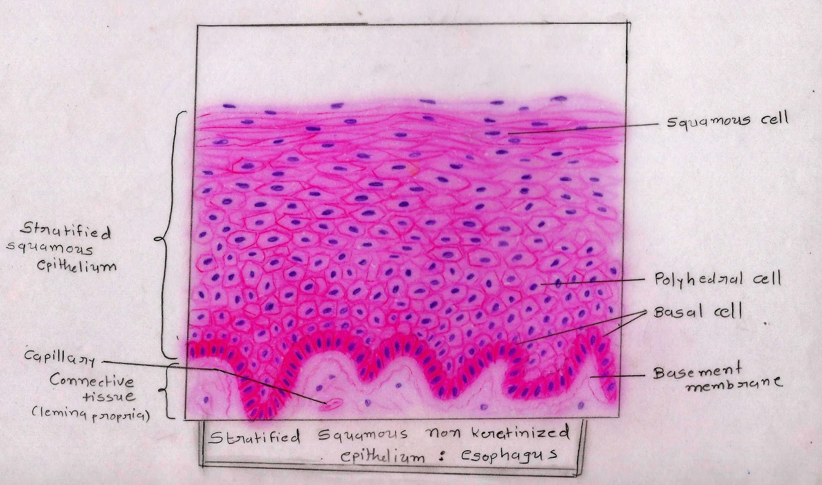
Stratified squamous, nonkeratinized epithelium stratified epithelia have multiple layers and are further classified by the shape of luminal cells. Cell biology & histology a560. Stratified epithelia are classified by the shape of the surface layer of cells.
nonkeratinized stratified squamous epithelium YouTube
This epithelium covers internal body surfaces which are exposed to some degree of physical trauma.
Learn vocabulary, terms, and more with flashcards, games, and other study tools.
Name the specific tissue at the pointer. The tissue is classified by the number of cell layers it has (simple=1 cell layer, stratified=more than 1 cell layer) and the shape of the cells (squamous=flat,. This is also stratified squamous epithelium, but notice the layer of keratin on the surface. This epithelium has 40 to 50 layers of cells.
It can be found in the lip, tongue, vagina, anal canal, esophagus, and palate(as in this case, soft palate specifically).
(5) lining lower 1/3 of anus. When living, nucleated cells are seen at the surface; The arrow indicates one of these squamous cells. Once again, there are many layers of cells but the surface layer is flattened, squamous cells.
It provides protection from abrasion and is keratinized on the external surface of the body.
The multiple layers of stratified squamous moist epithelium provide protection against friction and trauma to organs within the body. Where is this tissue type found? Histology slide courtesy of mt. Stratified squamous, nonkeratinized epithelium this image demonstrates the transition of epithelial cells from cuboidal at the basement membrane to squamous cells at the surface.
A layer of keratin has not formed;
Other sets by this creator. Name the specific tissue at the pointer. Cuboidal cells in the basal layers, round cells in the middle layers, and flattened (squamous) in the upper layers. The cells in this tissue are not all squamous (flat).
In this epithelium, the surface cells are living and possess a variety of organelles, as well as a prominent nucleus.
Thick skin only occurs on the palmar and plantar surfaces hands and feet, whereas thin skin occurs on all other parts of the body. It is found in the bladder, ureters and kidney. This is an example of nonkeratinized stratified squamous epithelium. The flat cells shown on the slide form several layers that make up this type of epithelial tissue.
It protects the tissue from damage and water loss.
Stratified squamous epithelium has multiple layers of cells becoming flattened as they move from the basal layer to the apical layers. And the epithelium remains moist. It is found in moist cavities subject to trauma, whereas the keratinized type is found on dry surfaces. Stratified squamous epithelium this is an audio histology slide!
It is named for the shape of the cells on the surface of the tissue.
Epithelial tissue covers or lines body surfaces as well as serving to absorb, filtrate, protect, and secrete various substances. Histology slide courtesy of william l. (2) lining of esophagus, (3) lining distal portion of urethra; At concordia college, moorhead, minnesota.
This epithelium shows a basal layer of cuboidal cells, then several layers of polygonal cells that become progressively more flat, until they become squamous at the luminal surface.
There is an absence of nuclei in the uppermost layers as these are dead cells, full of keratin. They change shape as they migrate from the basal layer to surface: Name the specific tissue at the pointer. The epithelial cells form between 10 and 20 layers.
Click on the button below the histology image and you will hear me.
Thick skin is covered by a stratified squamous keratinized epithelium.


