Identification points of general histology slides: Epithelial tissue covers or lines body surfaces as well as serving to absorb, filtrate, protect, and secrete various substances. Single layer of cells cube like cells rounded central nucleus 3:
Illustrations Stratified Squamous Keratinized Epithelium
They change shape as they migrate from the basal layer to surface:
Thick skin only occurs on the palmar and plantar surfaces hands and feet, whereas thin skin occurs on all other parts of the body.
Slide 114m (lip, monkey, h&e) view virtual slide. Transitional epithelium form of stratified epithelium. They have no nucleus or organelles. There is an absence of nuclei in the uppermost layers as these are dead cells, full of keratin.
Note here that the superficial layers are very thin and lack nuclei or any other structure in cells.
These layers, composed of keratin, form as the cells differentiate and die. The flat cells shown on the slide form several layers that make up this type of epithelial tissue. Single layer of cells tall cells oval nucleus oriented along the length of cell 4: Where is this tissue type found?
Thick skin is covered by a stratified squamous keratinized epithelium.
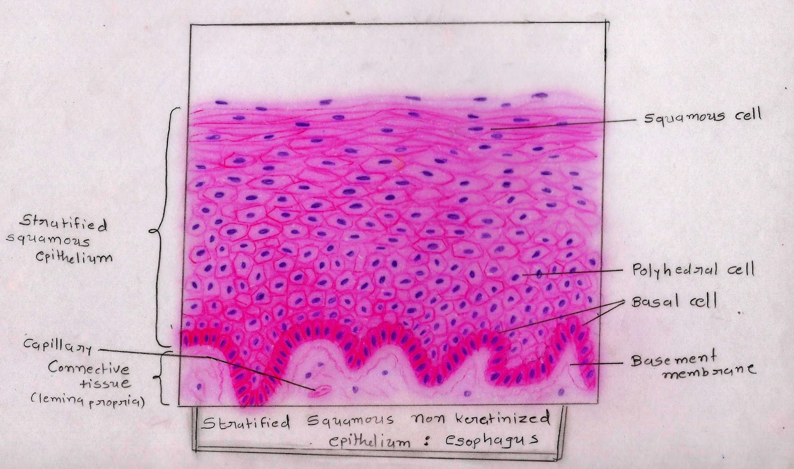
(2) lining of esophagus, (3) lining distal portion of urethra; Highly specialized to accommodate a great degree of stretch. At concordia college, moorhead, minnesota. Observe the thick cornified layer of stratified squamous keratinizing epithelium of the skin.
The tissue is classified by the number of cell layers it has (simple=1 cell layer, stratified=more than 1 cell layer) and the shape of the cells (squamous=flat,.
Click the button below to reveal the answer key: Cells which have been sloughed are indicated by the blue arrows. Stratified squamous nonkeratinizing epithelium lines the lumen of the esophagus. Thick skin only occurs on the palmar and plantar surfaces of hands and feet, whereas thin skin occurs on all other parts of the body.
This type of epithelium is found in mucous membranes.
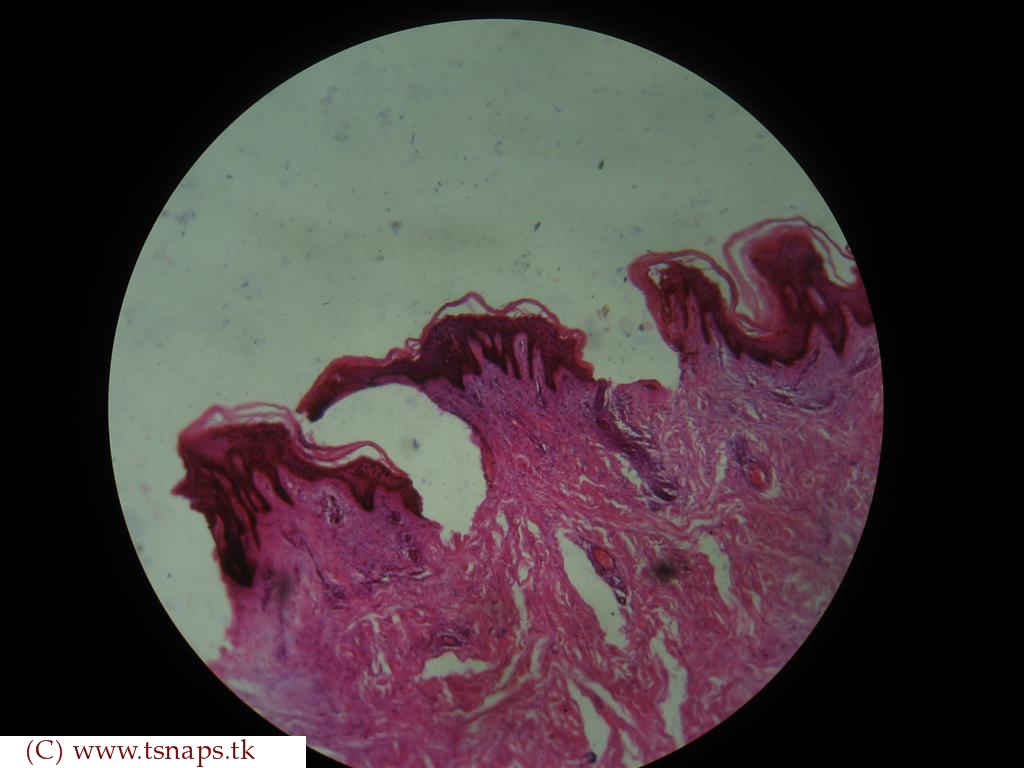
Stratified squamous keratinized epithelium 400x (palmar skin) the cells on the surface of stratified squamous keratinized epithelium are very flat. Name the specific tissue at the pointer. The epidermis of the skin is very thin and lines with the keratinized stratified squamous epithelium. The basal portion is composed of viable cells, while the outer layer is composed of dead cells made up almost entirely of the protein, keratin.
You might find out the dermal papillae, sebaceous glands, sweat glands, arrector pili muscles from the skin histology slide.
The epithelial cells form between 10 and 20 layers. Only a few layers of epithelial cells. Cuboidal cells in the basal layers, round cells in the middle layers, and flattened (squamous) in the upper layers. Not only are they flat, but they are no longer alive.
(5) lining lower 1/3 of anus.
The most superficial layers are only loosely attached and are steadily. Name the specific tissue at the pointer. So named because it has some features which are intermediate (transitional) between stratified cuboidal and stratified squamous epithelia. Histology slide courtesy of mt.
Stratified epithelia are classified by the shape of the surface.
Single layer of cells flat cells flat nucleus 2: Histology slide courtesy of william l. This epithelium has 40 to 50 layers of cells. Name the specific tissue at the pointer.
Epithelial tissue histology slide with microscopic images and labeled diagrams.
They are filled with a protein called keratin, which is what makes our skin waterproof. Note the changes in cellular morphology and intercellular spaces as one moves from the basal cells. It is found in the bladder, ureters and kidney. Slide 43 skin, sole of foot.
This is also stratified squamous epithelium, but notice the layer of keratin on the surface.
Stratified squamous, keratinized epithelium this epithelium, a subdivision of skin, covers the exterior, or dry, surface of the body that is exposed to the external environment.