• draw a color picture of a keratinized stratified squamous epithelium from thick skin. Biology q&a library this is astratified squamous epithelium keratinized. Epithelial tissues are those that encase the bodies of animals, both internally and externally.
Keratinized vs. Nonkerantinized Stratified Squamous
As the most important difference between the simple epithelium and the stratified epithelium is the number of the layer of cells, the functions of these layers also.
View 20210830_204054_2.jpg from bio 2050 at prince george's community college, largo.
A defining feature of this type of tissue is that one surface of the. If we just want to look at stratified squamous keratinized epithelium, we look at skin from one of the. The surface layer of it consists of dead cells. Stratified squamous, nonkeratinized epithelium this image demonstrates the transition of epithelial cells from cuboidal at the basement membrane to squamous cells at the surface.
Stratified keratinized epithelium is typically observed in the epidermis of land vertebrates, but it is also found in the papillae of the tongue, oral palate and esophagus of some animals eating hard food.
Label the parts indicated on the checklist. It is named for the shape of the cells on the surface of the tissue. Learn vocabulary, terms, and more with flashcards, games, and other study tools. Stratified squamous epithelium drawing :
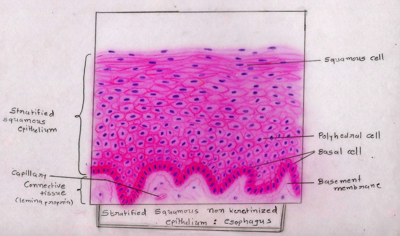
Ectoderm lamina propria skin covers the external surface.
Simple cuboidal epithelium total magnification: Stratified squamous epithelia are tissues formed from multiple layers of cells resting on a basement membrane, with the superficial layer(s) consisting of squamous cells. The cells in this tissue are not all squamous (flat). (palmar skin) although stratified squamous keratinized epithelium covers the entire surface of the body, most of it also includes hair, which makes the basic tissue structure harder to see.
Keratinized epithelium forms an effective barrier.
Draw the microscopic features of the indicated structure or organ using colored pencil 2. Keratinized stratified nuclei epidermis squamous epithelium keratinocytes dermis stratum stratum stratum corneum granulosum spinosum stratum basale free space Laboratory exercise 7 epithelial tissue instructions: This is due to the convention of.
Keratin is deposited on the surface.
Keratinized epithelium is a stratified squamous epithelium found in skin, epidermis of the palm of the hand and sole of the foot and the masticatory mucosa. Touch device users, explore by touch or with swipe gestures. A typical example of stratified squamous keratinized epithelium is the epidermis. Underlying cell layers can be made of cuboidal or columnar cells as well.
Examples of epithelial tissues include the cells that make up the external layer of the human skin and the stomach lining.
Keratinized epithelium may cause hardening of the cells of the stomach lining. In the deep layer of stratified squamous epithelium, you will find the columnar epithelium that rests on the basement membrane. Notice how the simple columnar cells lining the uterine cervix transitions to stratified squamous epithelium. Start studying epithelial tissue images and drawings.
Draw a color picture of a keratinized stratified squamous epithelium from thick skin.
The strata of the epidermis can be clearly observed in the image above, which is from thick skin of a mouse. Draw diagram of each type of epithelial tissue. Use the information in the passage to identify each of the cell types shown in the drawings. Passage of materials by diffusion and filtration.
Use slides 1 & 2 from the chapter 4 slides for inspiration.
The multiple layers of stratified squamous moist epithelium provide protection against friction and trauma to organs within the body. Squamous epithelium, non‐keratinized (blue arrow). The other layers adhere to one another to maintain structural integrity. This is astratified squamous epithelium keratinized.
Cute hand drawn vector illustration.
Many layers of nuclei can be seen and the cells nearest the free surface are squamous in appearance. Ppt simple squamous epithelium powerpoint presentation free download id 258513 /. Stratified squamous keratinized epithelium 40x. (keratinised stratified squamous epithelium) origin:
Only one layer is in contact with the basement membrane;
Mesoderm lumen generalised section epithelium of the body connective tissue beneath epithelium connective tissue, muscle, glands, etc dermis mesentery lining epithelium The arrow indicates one of these squamous cells. When autocomplete results are available use up and down arrows to review and enter to select. Each papilla is surrounded by a deep sulcus that receives ducts of the serous glands of von ebner.
Stratified squamous epithelium (keratinized) specimen:
Learn to draw stratified squamous keratinized epithelium histology diagram ( for mbbs and bds students) Although this epithelium is referred to as squamous, many cells within the layers may not be flattened; The stratified squamous epithelium consists of several layers of cells, where the cells in the apical layer and several layers present deep to it are squamous, but the cells in deeper layers vary from cuboidal to columnar. Blood vessel lined by endothelium (simple squamous epithelium) origin:
• label it with the following terms:
Functions of stratified squamous epithelia A stratified squamous epithelium consists of squamous epithelial cells arranged in layers upon a basal membrane. Eight to twelve circumvallate papillae are located along the sulcus terminalis, separating the anterior from the posterior portion of the tongue.