Under a microscope, epithelial cells are readily distinguished by the following features: Ppt simple squamous epithelium powerpoint presentation free download id 258513 /. The function of stratified epithelium is mainly protection.
Simple Squamous Epithelium Labeled Diagram slidedocnow
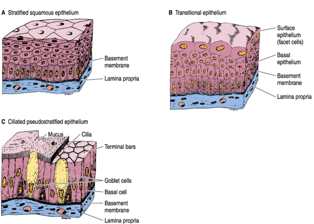
The stratified squamous epithelium consists of several layers of cells, where the cells in the apical layer and several layers present deep to it are squamous, but the cells in deeper layers vary from cuboidal to columnar.
Are branched forms of microvilli.
Solution for gastroesophageal juntion 10x draw and label stratified squamous , epithelium , of esophagus gastroesopheal juntion simple columar epithelium (of… As the most important difference between the simple epithelium and the stratified epithelium is the number of the layer of cells, the functions of. Layer that is exposed to the outside of the cell. A stratified epithelium consists of multiple stacked layers of cells.
This video describes how to draw stratified squamous non keratinized epithelium histology diagram.
X papilla dense irregular connective tissue 4. The cells in this tissue are not all squamous (flat). Cells in the deep layers are basically cuboidal but become more and more flat as they are pushed toward. In usual slides the boundaries between epithelial cells are often not clearly seen but because of the shape and spacing of the nuclei, the epithelium can be identified.
Draw and label the stratified squamous epithelium.
Draw a section showing the papillae. The pseudostratified columnar epithelium lining the. Secretion from the closely associated glands lubricates the surface of the nonkeratinized epithelium. Rumen histology slide identification points
Label the drawing with stratified squamous epithelium, dense regular connectives soe, papilla, magnification, and the name of the organ.
I hope the rumen slide labeled diagram might help you find out the structures mentioned above easily. In the stratified squamous epithelium labeled diagram, i tried to show you the columnar cells, elongated nucleus, and basement membrane. The arrow indicates one of these squamous cells. This type of epithelium is protective against chemical and mechanical damage, and water loss, and is found in skin, and oral epithelia.
Stratified squamous epithelium is the most common type of stratified epithelium in the human body.
Simple squamous epithelium is a single layer of platelike cells. Epithelium is a tissue that lines the internal surface of the body, as well as the internal organs. Is adapted to withstand abrasive forces c. It's difficult to see the basal lamina in the region of the dividing cells, in the basal layer.
This epithelium protects against physical and chemical damage.
Stratified squamous diagram photo of endothelial cells. The stratified epithelium is named by the shape of the most apical layer of cells, closest to the free space. The keratinization, or lack thereof, of the apical surface domains of the cells. (keratinised stratified squamous epithelium) origin:
Blood vessel lined by endothelium (simple squamous epithelium) origin:
Underlying cell layers can be made of cuboidal or columnar cells as well. This is an example of thin skin. Stratum corneum cell layers (variable thickness) and others; It is named for the shape of the cells on the surface of the tissue.
Is anchored on its basal surface to a basement membrane e.
The stratum basale (also known as stratum germinativum) is the inner layer of the epidermis. In the deepest layer new cells are produced by the division of stem cells. For figure with kidneys and drawing of stratified cuboidal Ectoderm lamina propria skin covers the external surface.
Form a brush border in.
Pseudostratified columnar epithelium is a single layer of ciliated, irregularly shaped cells containing many goblet cells. Passage of materials by diffusion and filtration. Cells are gradually pushed toward the surface by the production of newer cells. Bodytomy provides a labeled diagram to help you understand the structure and simple columnar epithelium:
A stratified squamous epithelium is made up of a number of layers and the cells of the outer layers are flat (squamous).
Stratified squamous keratinized epithelium 40x (palmar skin) although stratified squamous keratinized epithelium covers the entire surface of the body, most of it also includes hair, which makes the basic tissue structure harder to see. Stratified squamous epithelium (keratinized) lining in the mucosa or papillae of rumen slide. This type of epithelium is thin and leaky, allowing for diffuse to occur. Layers at the surface where keratin cements the debris of dead squamous cells, the non living keratin layer.
Cute hand drawn vector illustration.
Stratified squamous in the oral cavity simple squamous in the lungs simple columnar in the digestive tract image 3: Mesoderm lumen generalised section epithelium of the body connective tissue beneath epithelium connective tissue, muscle, glands, etc dermis mesentery lining epithelium A typical example of stratified squamous keratinized epithelium is the epidermis. Layer where cells are continually dividing by mitosis.
Stratified squamous epithelium is multilayered, which would impede diffusion.
If we just want to look at stratified squamous keratinized epithelium, we look at skin from one of the few. Increase the magnification to 400x at this magnification, the more cuboidal nature of the bottom layer should be visible. In fact, this specific role is reflected in the direct influence of. Remember, the circle represents your field of view, so your drawing should be in proportion.
Thus as gas and nutrients are exchanged through diffusion, simple squamous is the best choice.
Microscopic images of the different types of epithelium modified cc by 2.0 by smile with your eyes url: Stratified squamous epithelium drawing :