Ppt simple squamous epithelium powerpoint presentation free download id 258513 /. Tap card to see definition 👆. A defining feature of this type of tissue is that one surface of the.
PPT Simple Squamous Epithelium PowerPoint Presentation
In the stratified squamous epithelium labeled diagram, i tried to show you the columnar cells, elongated nucleus, and basement membrane.
The esophageal squamous epithelium is nonkeratinizing, i.e., it.
Secretion from the closely associated glands lubricates the surface of the nonkeratinized epithelium. Hence, simple squamous epithelia is located in the alveolar sacs of the lungs and in the endothelium of. Each papilla is surrounded by a deep sulcus that receives ducts of the serous glands of von ebner. Bottom layer of cells in this epithelium.
Draw a color picture of a keratinized stratified squamous epithelium from thick skin.
Clear area between living and. Keratinized epithelium forms an effective barrier. Stratified squamous keratinized epithelium 400x (palmar skin) the cells on the surface of stratified squamous keratinized epithelium are very flat. The strata of the epidermis can be clearly observed in the image above, which is from thick.
Keratin is deposited on the surface.
Blood vessel lined by endothelium (simple squamous epithelium) origin: Keratinized stratified squamous epithelium eight to twelve circumvallate papillae are located along the sulcus terminalis, separating the anterior from the posterior portion of the tongue. Learn to draw stratified squamous keratinized epithelium histology diagram ( for mbbs and bds students) It is named for the shape of the cells on the surface of the tissue.
As the most important difference between the simple epithelium and the stratified epithelium is the number of the layer of cells, the functions of these layers also.
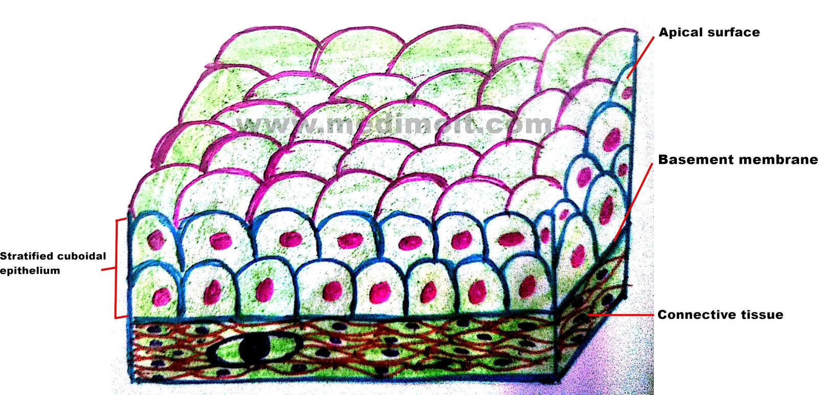
• label it with the following terms: The other layers adhere to one another to maintain structural integrity. A stratified squamous epithelium consists of squamous epithelial cells arranged in layers upon a basal membrane. Draw diagram of each type of epithelial tissue.
The parts of the mouth that feel a little rough such as the upper surface of the tongue and the hard palate at the roof of the mouth contain keratinized epithelia.
View 20210830_204054_2.jpg from bio 2050 at prince george's community college, largo. (keratinised stratified squamous epithelium) origin: The stratified squamous epithelium consists of several layers of cells, where the cells in the apical layer and several layers present deep to it are squamous, but the cells in deeper layers vary from cuboidal to columnar. The surface layer of it consists of dead cells.
Write that the thin cells of squamous epithelia provide an ideal surface for diffusion and filtration;
Cute hand drawn vector illustration. Use slides 1 & 2 from the chapter 4 slides for inspiration. They are filled with a protein called keratin, which is what makes our skin waterproof. A typical example of stratified squamous keratinized epithelium is the epidermis.
The arrow indicates one of these squamous cells.
Protein that makes this epithelium water tight. Keratinized epithelium may cause hardening of the cells of the stomach lining. Keratinized stratified nuclei epidermis squamous epithelium keratinocytes dermis stratum stratum stratum corneum granulosum spinosum stratum basale free space Laboratory exercise 7 epithelial tissue instructions:
Although this epithelium is referred to as squamous, many cells within the layers may not be flattened;
Ectoderm lamina propria skin covers the external surface. Stratified squamous epithelium drawing : Simple cuboidal epithelium total magnification: This is due to the convention of.
Multiple layers of flattened cells, basal cells typically are cuboidal, while apical cells are squamous.
The cells in this tissue are not all squamous (flat). Not only are they flat, but they are no longer alive. Click card to see definition 👆. This image demonstrates the transition of epithelial cells from cuboidal at the basement membrane to squamous cells at the surface.
Use slides 1 & 2 from the
In a stratified squamous moist epithelium, cells retain their nuclei, even at the surface (blue arrows). Passage of materials by diffusion and filtration. Mesoderm lumen generalised section epithelium of the body connective tissue beneath epithelium connective tissue, muscle, glands, etc dermis mesentery lining epithelium The basal layer, the spinous layer, the.
What cellular junction accounts for the spiny looking cell in the second layer?
Only one layer is in contact with the basement membrane; Epithelial tissues are those that encase the bodies of animals, both internally and externally. These tissues are formed by four layers: Surface cells are sloughed off into the lumen (black arrows).
• draw a color picture of a keratinized stratified squamous epithelium from thick skin.
They have no nucleus or organelles. Stratified squamous epithelium (keratinized) specimen: Where is keratinized stratified squamous epithelium found in the body? Surface cells are alive and kept moist.
Keratinized epithelium is a stratified squamous epithelium found in skin, epidermis of the palm of the hand and sole of the foot and the masticatory mucosa.
Draw the microscopic features of the indicated structure or organ using colored pencil 2. Stratified keratinized epithelium is typically observed in the epidermis of land vertebrates, but it is also found in the papillae of the tongue, oral palate and esophagus of some animals eating hard food.