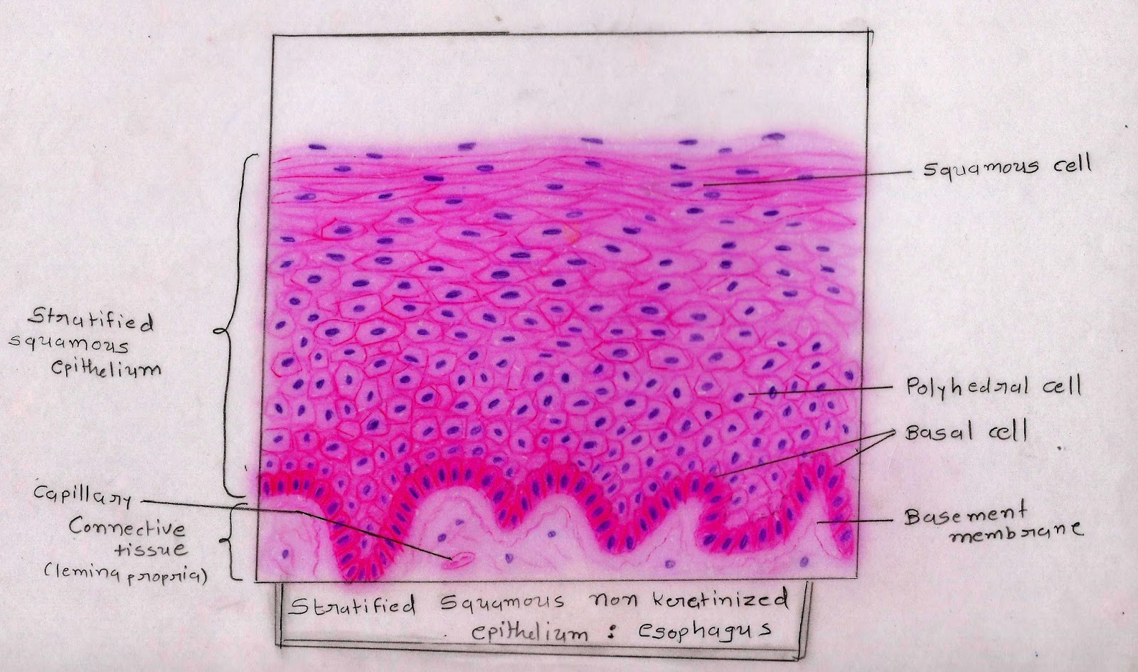
The stratified squamous epithelium consists of squamous (flattened) epithelial cells arranged in layers upon a basal membrane. Epithelial tissue histology slide with microscopic images and labeled diagrams. In stratified epithelia only the basal layer of cells touches the basal lamina.
Photomicrograph showing stratified squamous epithelium
Underlying cell layers can be made of cuboidal or columnar cells as well.
This is an example of thin skin.
This epithelium covers internal body surfaces which are exposed to some degree of physical trauma. This illustration is included in the following illustration toolkit. These labelled diagrams should closely follow the current science courses in histology, anatomy and embryology and complement the virtual microscopy used in the current medical course. The function of stratified epithelium is mainly protection.
(a) stratified squamous epithelium of the first esophageal region in.
This is a section of the skin clearly revealing the stratified squamous epithelium of the epidermis and the reticular layer in the more superficial aspect of the dermis. Stratified squamous epithelia have two or more layers of cells, with a superficial squamous layer and basal layers that are usually cuboidal or columnar. As the most important difference between the simple epithelium and the stratified epithelium is the number of the layer of cells, the functions of. They are sketches from selected slides used in class from the teaching slide set.
Stratified squamous epithelia are tissues formed from multiple layers of cells resting on a basement membrane, with the superficial layer(s) consisting of squamous cells.
Ectoderm lamina propria skin covers the external. Anatomic opening of the endocervical canal onto the ectocervix Simple squamous epithelium (diagram) stratified squamous epithelium (diagram) simple cuboidal epithelium (diagram) simple columnar epithelium (diagram) ciliated pseudostratified columnar. Notice the dead cornified layer of the epidermis at the surface pulling away from the living cell layers beneath it.
The muscularis mucosae is usually absent in the upper third.
What type of tissue is this? In fact, this specific role is reflected in the direct influence of. Terms in this set (50) simple epithelium. This epithelium contains 5 layers:
Stratified squamous keratinising epithelium this type of epithelium is protective against chemical and mechanical damage, and water loss, and is found in skin, and oral epithelia.
A typical example of stratified squamous keratinized epithelium is the epidermis. It is the shape of the cells in the top layer (free surface) that classifies the type of stratified epithelium. The size and shape of cells vary in the different layers. The stratified squamous epithelium consists of several layers of cells, where the cells in the apical layer and several layers present deep to it are squamous, but the cells in deeper layers vary from cuboidal to columnar.
The nuclei of these cells become condensed and eventually disappear as they reach the outermost.
These labelled diagrams should closely follow the current science courses in histology, anatomy and. Normally lined by nonkeratinizing stratified squamous epithelium endocervix (endocervical canal): A stratified squamous epithelium contains many sheets of cells, where the cells in the apical layer and several layers present deep to it are squamous, but the cells in deeper layers vary from cuboidal to columnar. Mucosa lined cylindrical canal leading from ectocervix to uterine cavity normally lined by single layer of mucinous columnar epithelium external os:
Powerpoint (win & mac compatible) price:
Mitosis occurs in this basal layer and the newly produced cells differentiate as they are forced outward. Functions of stratified squamous epithelia A thick stratified squamous nonkeratinized epithelium lines the esophagus. Keratinized stratified squamous epithelium is a type of stratified epithelium that contains numerous layers of squamous cells, called keratinocytes, in which the superficial layer of cells is keratinized.this type of epithelium comprises the epidermis of the skin.
What might be the function of keratin, and in what kinds of tissues would you expect to find stratified squamous keratinizing.
This epithelium shows a basal layer of cuboidal cells, then several layers of polygonal cells that become progressively more flat, until they become squamous at the luminal surface. Stratified squamous epithelium illustration figure drawing diagram image. The lamina propria underlying the epithelium possesses lymphoid structures and localized mucous glands (not shown here) in the lower third and sometimes in the upper third. This video describes how to draw stratified squamous non keratinized epithelium histology diagram.
The drawings of histology images were originally.
(keratinised stratified squamous epithelium) origin: I will show you everything about the rumen histology slide with their identification point and labeled diagram. The drawings of histology images were originally. The keratinization, or lack thereof, of the apical surface domains of the cells.
What type of tissue is this?
Stratified squamous with keratin in this image note the layers of keratin on the apical surface of the epithelium, superficial to the squamous cells. Click to see the original works with their full license. Contains the stem cells of the.


