Functions of stratified squamous epithelia This illustration is included in the following illustration toolkit. If we just want to look at stratified squamous keratinized epithelium, we look at skin from one of the few.
High magnification image of the keratinised stratified
This is due to the convention of.
Keratinized stratified nuclei epidermis squamous epithelium keratinocytes dermis stratum stratum stratum corneum granulosum spinosum stratum basale free space
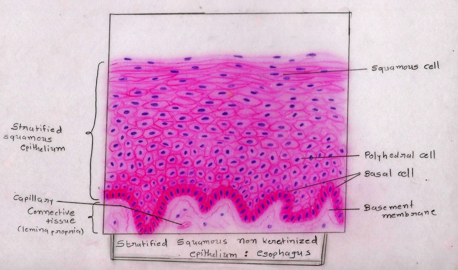
The cells in this tissue are not all squamous (flat). From your observations, do you think stratified squamous epithelium is the; Stratified squamous epithelium drawing : Stratified squamous epithelia are tissues formed from multiple layers of cells resting on a basement membrane, with the superficial layer(s) consisting of squamous cells.
The other layers adhere to one another to maintain structural integrity.
Learn to draw stratified squamous keratinized epithelium histology diagram ( for mbbs and bds students) A stratified squamous epithelium consists of squamous epithelial cells arranged in layers upon a basal membrane. Start studying epithelial tissue images and drawings. Blood vessel lined by endothelium (simple squamous epithelium) origin:
Discover millions of stock images, photos, video and audio.
Only one layer is in contact with the basement membrane; Stratified squamous, nonkeratinized epithelium this image demonstrates the transition of epithelial cells from cuboidal at the basement membrane to squamous cells at the surface. Cute hand drawn vector illustration. Although this epithelium is referred to as squamous, many cells within the layers may not be flattened;
In the deepest layer new cells are produced by the division of stem cells.
Brush border & terminal bars. Ppt simple squamous epithelium powerpoint presentation free download id 258513 /. • draw a color picture of a keratinized stratified squamous epithelium from thick skin. The pseudostratified columnar epithelium lining the.
Pseudostratified columnar epithelium is a single layer of ciliated, irregularly shaped cells containing many goblet cells.
As the most important difference between the simple epithelium and the stratified epithelium is the number of the layer of cells, the functions of these layers also. The stratified squamous epithelium consists of several layers of cells, where the cells in the apical layer and several layers present deep to it are squamous, but the cells in deeper layers vary from cuboidal to columnar. Stratified squamous keratinized epithelium 40x (palmar skin) although stratified squamous keratinized epithelium covers the entire surface of the body, most of it also includes hair, which makes the basic tissue structure harder to see. Stratified squamous in the oral cavity simple squamous in the lungs simple columnar in the digestive tract image 3:
Click on the button or title to get to the appropriate image:
Shop for stratified squamous epithelium wall art from the world's greatest living artists. Underlying cell layers can be made of cuboidal or columnar cells as well. In the deep layer of stratified squamous epithelium, you will find the columnar epithelium that rests on the basement membrane. It is named for the shape of the cells on the surface of the tissue.
(keratinised stratified squamous epithelium) origin:
Powerpoint (win & mac compatible) price: The multiple layers of stratified squamous moist epithelium provide protection against friction and trauma to organs within the body. Use slides 1 & 2 from the chapter 4 slides for inspiration. Cells are gradually pushed toward the surface by the production of newer cells.
Ectoderm lamina propria skin covers the external surface.
Microscopic images of the different types of epithelium modified cc by 2.0 by smile with your eyes url: In usual slides the boundaries between epithelial cells are often not clearly seen but because of the shape and spacing of the nuclei, the epithelium can be identified. The stratum basale (also known as stratum germinativum) is the inner layer of the epidermis. Stratified squamous epithelium illustration figure drawing diagram image.
• label it with the following terms:
A stratified squamous epithelium is made up of a number of layers and the cells of the outer layers are flat (squamous). Passage of materials by diffusion and filtration. Choose your favorite stratified squamous epithelium designs and purchase them as wall art, home decor, phone cases, tote bags, and more! The arrow indicates one of these squamous cells.
Compare your drawing of simple cuboidal epithelium from the previous activity to your sketch of stratified squamous epithelium in this activity.
Learn vocabulary, terms, and more with flashcards, games, and other study tools. Cells in the deep layers are basically cuboidal but become more and more flat as they are pushed toward.