The simple squamous epithelium is different from other types of epithelial tissue such as simple cuboidal, simple columnar, and stratified squamous epithelium in that it is only made of one layer. The lining of the mouth and vagina: Functions of stratified squamous epithelia
PPT Simple Squamous Epithelium PowerPoint Presentation
This epithelium contains 5 layers:
The function of stratified epithelium is mainly protection.
The keratinization, or lack thereof, of the apical surface domains of the cells. As the most important difference between the simple epithelium and the stratified epithelium is the number of the layer of cells, the functions of. Evaluation of the conventional method for. Powerpoint (win & mac compatible) price:
Stratified squamous diagram photo of endothelial cells.
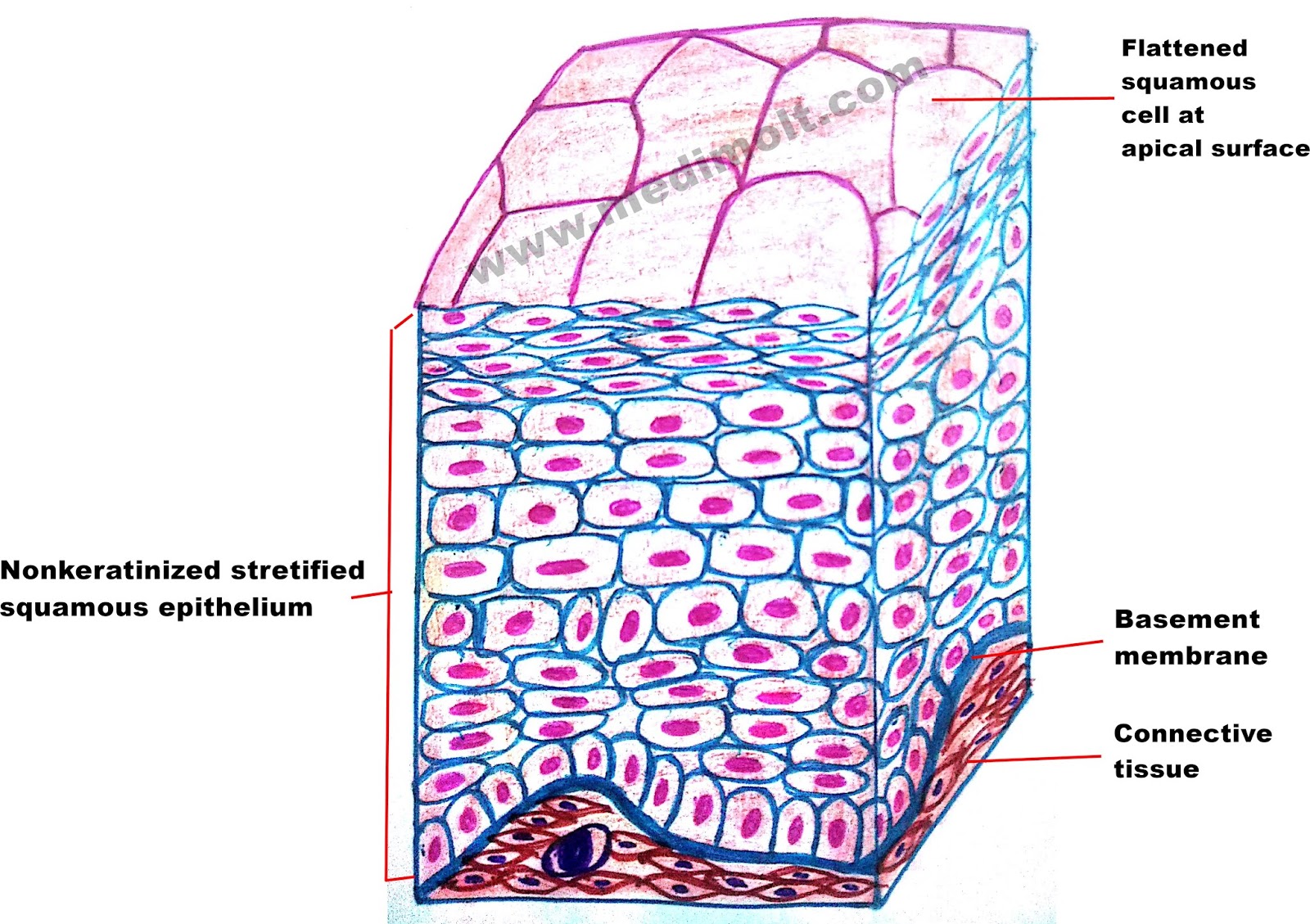
In fact, this specific role is reflected in the direct influence of. The oesophagus is an example of a stratified squamous non keratinising epithelium. Made up of several layers of cells, continuously sloughed off and regenerated. Stratified squamous epithelia are tissues formed from multiple layers of cells resting on a basement membrane, with the superficial layer(s) consisting of squamous cells.
A keratinized stratified squamous epithelium.
Learn to draw stratified squamous keratinized epithelium histology diagram ( for mbbs and bds students) Basale (sb) with melanocytes ( * ). The older layer of cells is pushed upwards and becomes flat. The stratum corneum varies in thickness from one to two cells to as many as ten to twenty cells.
3 separate models type of epithelium in body lining skin, esophagus, oral mucosa etc.
This video describes how to draw stratified squamous non keratinized epithelium histology diagram. The deepest layer is made up of columnar cells. The stratified squamous epithelium consists of cell layers in which the superficial layer consists of squamous epithelial cells while the underlying cell layers have various types of cells. Stratified squamous epithelium diagram and structure it has two or more layers of cells.
Bodytomy provides a labeled diagram to help you understand the structure and simple columnar epithelium:
Tissue diagram stratified squamous epithelium. There is a keratinized stratified squamous epithelium lining in the mucosal surface of the sample tissue. The outer layer of your skin is the epidermis: Download scientific diagram | keratinized stratified squamous epithelium.
A typical example of stratified squamous keratinized epithelium is the epidermis.
Identify the type of epithelium indicated by the diagram to the right: Underlying cell layers can be made of cuboidal or columnar cells as well. Keratinized stratified squamous epithelium is a type of stratified epithelium that contains numerous layers of squamous cells, called keratinocytes, in which the superficial layer of cells is keratinized.this type of epithelium comprises the epidermis of the skin. Contains the stem cells of the.
The dense collagen, reticular, and elastic fibers are present in the core of each papilla of the provided sample tissue.
This illustration is included in the following illustration toolkit. Identify the type of epithelium indicated by the black arrow: A typical example of stratified squamous keratinized epithelium is the epidermis. Epithelium / ˌ ɛ p ɪ ˈ θ iː l i ə m / is one of the four basic types of animal tissue, along with connective tissue, muscle tissue and nervous tissue.it is a thin, continuous, protective layer of compactly packed cells with little intercellular matrix.epithelial tissues line the outer surfaces of organs and blood vessels throughout the body, as well as the inner surfaces of cavities in.
Download scientific diagram | the nonkeratinized stratified squamous epithelium of the tongue dorsum displaying the str.
Stratified squamous epithelium illustration figure drawing diagram image. The stratified squamous epithelium consists of several layers of cells, where the cells in the apical layer and several layers present deep to it are squamous, but the cells in deeper layers vary from cuboidal to columnar. In the stratified squamous epithelium labeled diagram, i tried to show you the columnar cells, elongated nucleus, and basement membrane. Mammary glands, sweat gland and salivary glands
Epithelium is a tissue that lines the internal surface of the body, as well as the internal organs.
The lower layer is columnar and metabolically active: The cells in the apical layer and several layers deep to it are squamous while the cells in deeper layers vary from cuboidal to columnar. For the image and diagram below, label each layer of the epidermis and its cells:


:background_color(FFFFFF):format(jpeg)/images/library/2338/A4WBRjTb2LJJRtmn2wVxCw_stratified_squamous_epithelia02.png)

