What is the cell shape of this tissue? Simple squamous tissues / organs: Where are the locations of this tissue?
Non Keratinized Stratified Squamous Epithelium
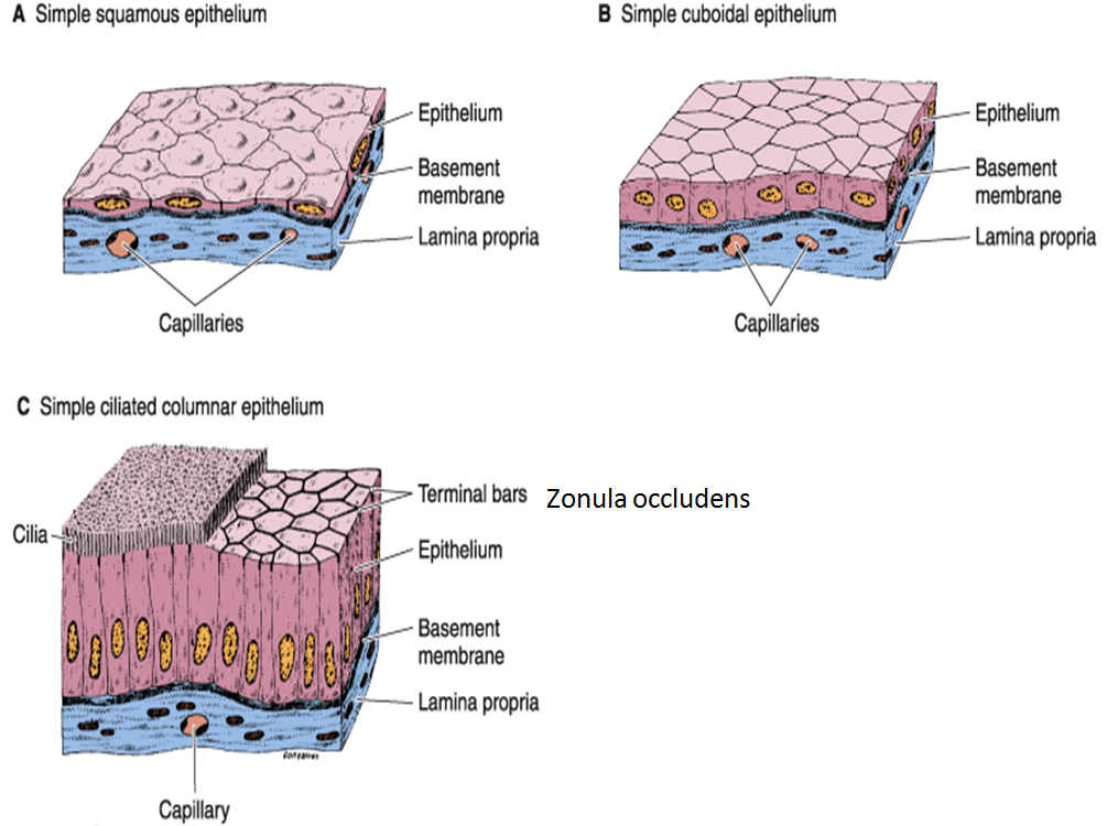
There are three types of epithelial cells which differ in their shape and function.
Stratified squamous diagram photo of endothelial cells.
The stratified squamous epithelium is divided into two classes based on the accumulation of keratin by the cells towards the surface; (keratinised stratified squamous epithelium) origin: Physical protection against abrasion, pathogens, and chemical attacks. Pseudostratified columnar (respiratory epithelium) more fully:
A typical example of stratified squamous keratinized epithelium is the epidermis.
The tunica muscularis of the rumen diagram showed inner circular and outer longitudinal layers of. The lower layer is columnar and metabolically active: Lining of the mouth, vagina. This epithelium contains 5 layers:
The lining of the mouth and vagina:
Learn about the structures, locations, and functions of this epithelial tissue, and then test yourself with labeled images, hints, and answer keys that put you in control. The oesophagus is an example of a stratified squamous non keratinising epithelium. Mammary glands, sweat gland and salivary glands Simple columnar tissue / organ:
Epithelium is a tissue that lines the internal surface of the body, as well as the internal organs.
As in the case of other stratified epithelium, the cells in the deeper layers might be different than the layer on the top. Pseudostratified ciliated columnar epithelium with goblet cells tissue / organ: The function of stratified epithelium is mainly protection. A typical example of stratified squamous keratinized epithelium is the epidermis.
1 endothelium lining blood vessel 2 mesothelium of serosa covering lung epithelium:
In fact, this specific role is. Layers at the surface where keratin cements the debris of dead squamous cells, the non living keratin layer. This type of epithelial tissue covers body parts that are exposed to frequent frictional forces or. The deepest layer is made up of columnar cells.
Keratinized stratified squamous epithelium is a type of stratified epithelium that contains numerous layers of squamous cells, called keratinocytes, in which the superficial layer of cells is keratinized.
The stratified epithelium is named by the shape of the most apical layer of cells, closest to the free space. A palentine tonsil b lingual الا pharyngeal d palentine e palentine tonsil. Anatomy and physiology questions and answers. These labelled diagrams should closely follow the current science courses in histology, anatomy and embryology and complement the virtual microscopy.
If we just want to look at stratified squamous keratinized epithelium, we look at skin from one of the few.
The stratified squamous epithelium consists of cell layers in which the superficial layer consists of squamous epithelial cells while the underlying cell layers have various types of cells. The outer layer of your skin is the epidermis: The rumen slide labeled diagrams represent the tunica mucosa with the lining epithelium and papillae. A stratified epithelium consists of multiple stacked layers of cells.
This video describes how to draw stratified squamous non keratinized epithelium histology diagram.
In addition, the diagram showed the other three layers of the rumen tissue. The older layer of cells is pushed upwards and becomes flat. Tissue diagram stratified squamous epithelium. A keratinized stratified squamous epithelium.
Throat, skin surface (keratinized), esophagus, rectum, and anus.
Learn to draw stratified squamous keratinized epithelium histology diagram ( for mbbs and bds students) Bodytomy provides a labeled diagram to help you understand the structure and simple columnar epithelium: This type of epithelium comprises the epidermis of the skin. For the image and diagram below, label each layer of the epidermis and its cells:
In the stratified squamous epithelium labeled diagram, i tried to show you the columnar cells, elongated nucleus, and basement membrane.
Stratified squamous epithelium is the most common type of stratified epithelium in the human body. Stratified squamous keratinized epithelium 40x (palmar skin) although stratified squamous keratinized epithelium covers the entire surface of the body, most of it also includes hair, which makes the basic tissue structure harder to see. This epithelium protects against physical and chemical damage. Made up of several layers of cells, continuously sloughed off and regenerated.
Underlying cell layers can be made of cuboidal or columnar cells as well.
The stratified squamous epithelium consists of several layers of cells, where the cells in the apical layer and several layers present deep to it are squamous, but the cells in deeper layers vary from cuboidal to columnar. Ectoderm lamina propria skin covers the external surface. Layer where cells are continually dividing by mitosis. Classification of stratified squamous epithelium.
What are the functions of this tissue?
Layer that is exposed to the outside of the cell. Thin, flat, and irregular in shape. The simple squamous epithelium is different from other types of epithelial tissue such as simple cuboidal, simple columnar, and stratified squamous epithelium in that it is only made of one layer. Using the diagram below, label the correct e d 2 b (b) histology of palatine tonsil crypt stratified squamous epithelium germinal centers tonsil indicated by the letter.
The stratified columnar epithelium has multiple layers of cells in which the apical layer is made up of columnar cells while the deeper layer can be either cuboidal or columnar.
As the most important difference between the simple epithelium and the stratified epithelium is the number of the layer of cells, the functions of.