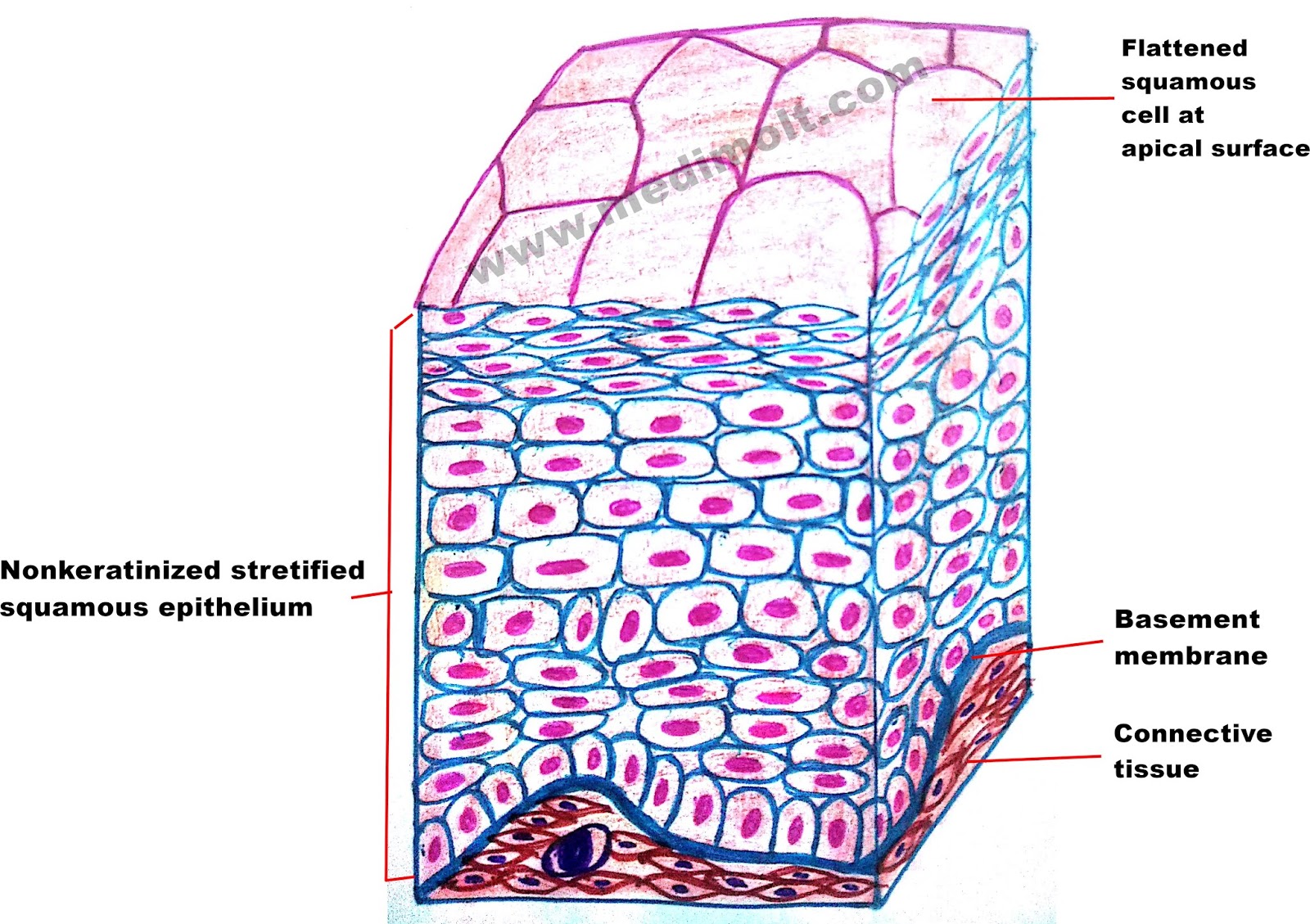
Stratified squamous epithelium is the most common type of stratified epithelium in the human body. Keratinized type forms the epidermis of the skin, a dry membrane. The function of stratified epithelium is mainly protection.
Illustration Of Stratified Squamous Photograph by Science
Epithelial tissues are of following types:
Reset help stratified squamous ophold group 2 basional epitel connective liste grove group 2 basement membrane group nude of the bols group group part a drag the labels onto the diagram to identify the tissues and structures.
Cuboidal or columnar cells may be seen in the deeper layers. Epithelial tissue histology slide with microscopic images and labeled diagrams. Some stratified squamous epithelia are extensively keratinized, whereas others are keratinized either minimally or not at all. A typical example of stratified squamous keratinized epithelium is the epidermis.
The cells gradually become larger and more squamous as the cells migrate from the.
This type of epithelial tissue covers body parts that are exposed to frequent frictional forces or. The outline of the cells is slightly irregular wherein the cells fit in forming a lining or covering. The deepest layer is made up of columnar cells. In fact, this specific role is reflected in the direct influence of.
Epithelium is a tissue that lines the internal surface of the body, as well as the internal organs.
As the most important difference between the simple epithelium and the stratified epithelium is the number of the layer of cells, the functions of. Tissue diagram stratified squamous epithelium. (1) simple squamous epithelium (2) stratified squamous epithelium (3) columnar epithelium, and (4) cubodial epithelium. The oesophagus is an example of a stratified squamous non keratinising epithelium.
The keratinization, or lack thereof, of the apical surface domains of the cells.
The simple squamous epithelium is different from other types of epithelial tissue such as simple cuboidal, simple columnar, and stratified squamous epithelium in that it is only made of one layer. There are three types of epithelial cells which differ in their shape and function. Stratified squamous epithelium thick membrane composed of several cell layers; Under a microscope, epithelial cells are readily distinguished by the following features:
On the bssis of shape / structural modifications of cells.
The cells in the apical layer and several layers deep to it are squamous while the cells in deeper layers vary from cuboidal to columnar. Diagram of a nonkeratinizing stratified squamous epithelium a stratified squamous epithelium consists of more than one (typically many) layers, or strata, of epithelial cells (see figure). Learn vocabulary, terms, and more with flashcards, games, and other study tools. There are three types of epithelial cells, which differ in their shape and function.
Epithelial tissue study guide by ebonie_williams6 includes 12 questions covering vocabulary, terms and more.
The stratified squamous epithelium consists of cell layers in which the superficial layer consists of squamous epithelial cells while the underlying cell layers have various types of cells. The simple epithelia (cells arranged in a single layer) promote the diffusion in tissues namely the regions of gas exchange in the lungs and the exchange of the wastes and nutrients at the blood capillaries. The stratified squamous epithelium consists of several layers of cells, where the cells in the apical layer and several layers present deep to it are squamous, but the cells in deeper layers vary from cuboidal to columnar. Nonkeratinized type forms the moist lining of the esophagus, mouth, and vagina;
02 part a drag the labels onto the diagram to identity the tissues and structures.
Learn vocabulary, terms, and more with flashcards, games, and other study tools. Basal cells are active in mitosis and produced the cells of the more superficial layers. Functions as lining for body cavities, ducts and tubes. Stratified squamous epithelia are tissues formed from multiple layers of cells resting on a basement membrane, with the superficial layer(s) consisting of squamous cells.
Basal cells are cuboidal or columnar and metabolically active;
The top layer may be covered with dead cells containing keratin. Quizlet flashcards, activities and games help you improve your grades. The skin is an example of a keratinized, stratified squamous epithelium. A typical example of stratified squamous keratinized epithelium is the epidermis.
These tissues differ in structure that correlate with their unique functions.
Composed of single layer of cells. Stratified squamous diagram photo of endothelial cells. (a) simple squamous epithelial tissue. Bodytomy provides a labeled diagram to help you understand the structure and simple columnar epithelium:
Functions of stratified squamous epithelia
Surface cells are flattened (squamous) in the kera tinized type, the surface cells are full of keratin and dead; The apical cells appear squamous, whereas the basal layer contains either columnar or cuboidal cells. Protects underlying tissues in areas subject to abrasion location:


