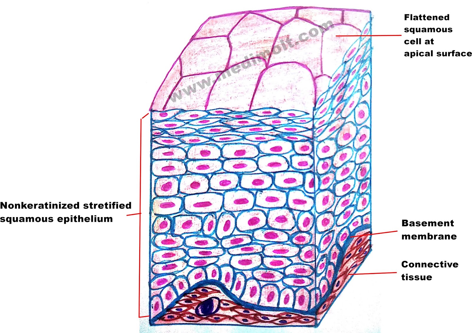
The modification of the cells on the apical surface is based on the location and function of. Stratified squamous epithelium illustration figure drawing diagram image. Stratified cuboidal epithelium allows glands and organs to create a separation between the functioning cells of organ or gland and the vessels that feed it.
Types Of Epithelium Stock Illustration Download Image
This type of epithelium is protective against chemical and mechanical damage, and water loss, and is found in skin, and oral epithelia.
The stratified columnar epithelium has multiple layers of cells in which the apical layer is made up of columnar cells while the deeper layer can be either cuboidal or columnar.
This is not a true stratified epithelium but appears to be stratified. Mesoderm lumen generalised section epithelium of the body connective tissue beneath epithelium connective tissue, muscle, glands, etc dermis mesentery lining epithelium A typical example of stratified squamous keratinized epithelium is the epidermis. Huge collection, amazing choice, 100+ million high quality, affordable rf and rm images.
Surface cells are sloughed off into the lumen (black arrows).
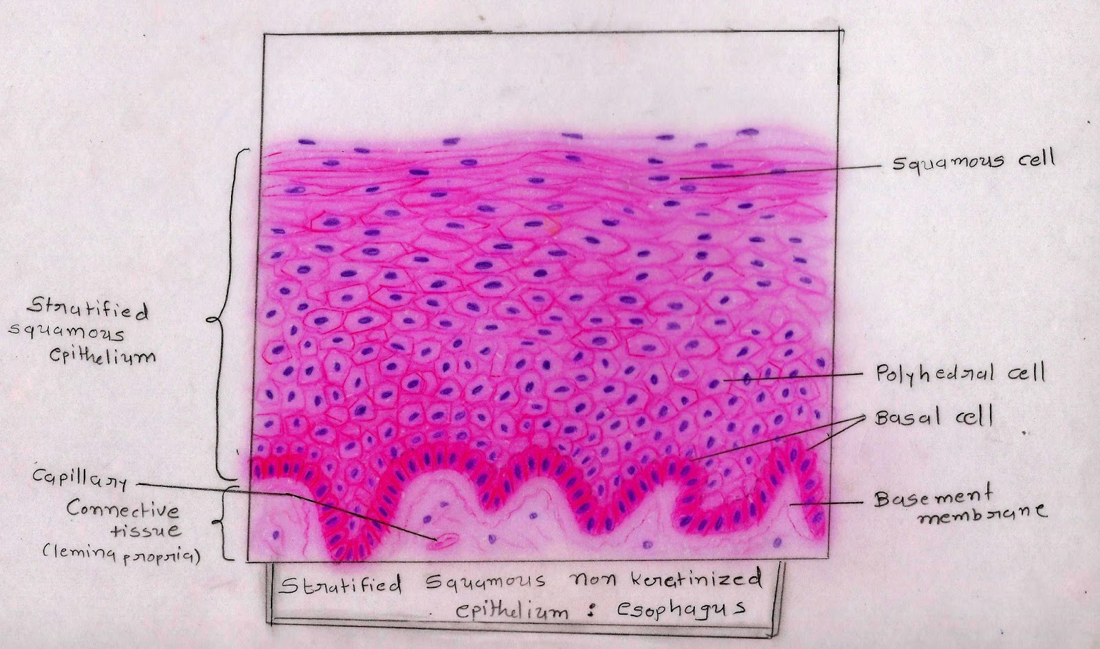
Identify the variant subtype of stratified epithelium present in the specimens provided. In a stratified squamous moist epithelium, cells retain their nuclei, even at the surface (blue arrows). Powerpoint (win & mac compatible) price: Note the difference in the appearance/morphology of the cells seen on the outer free surface of each variety.
Bodytomy provides a labeled diagram to help you understand the structure and simple columnar epithelium:
Stratified columnar epithelium is a type of epithelial tissue composed of two or more layers of columnar epithelial cells.epithelial tissue is one of the body’s primary tissues and lines the surfaces and insides of structures in many parts of the body. Stratified squamous diagram photo of endothelial cells. This image demonstrates the transition of epithelial cells from cuboidal at the basement membrane to squamous cells at the surface. In the circle below, draw a representative sample of the epithelial cells, taking care to correctly and clearly draw their true shape in the slide and an accurate number of layers if it is a stratified epithelium.
It's difficult to see the basal lamina in the region of the dividing cells, in the basal layer.
The keratinization, or lack thereof, of the apical surface domains of the cells. Note the dome shaped appearance of the surface layer of epithelial cells (a.k.a umbrella cells, blue arrow) and prominent oval shaped Epithelium is a tissue that lines the internal surface of the. • draw or paste the image of the varieties of stratified epithelium as observed under hpo.
A typical example of stratified squamous keratinized epithelium is the epidermis.
Underlying cell layers can be made of cuboidal or columnar cells as well. Structure of stratified cuboidal epithelium. The cells are tightly packed to ensure no gap is present in two cells. (keratinised stratified squamous epithelium) origin:
As in the case of other stratified epithelium, the cells in the deeper layers might be different than the layer on the top.
The basal layer of the epithelium is attached to the basement membrane. This is a stratified epithelium, multiple cell layers are visible. The layers of squamous epithelial cells beneath the cuboidal cells replace damaged cells as needed to maintain the epithelial lining. In fact, this specific role is reflected in the direct influence of.
The function of stratified epithelium is mainly protection.
Simple squamous epithelium under microscope drawing. Two or more layers of cells indicate the tissue has strata (layers), and are called stratified. A stratified squamous epithelium is a tissue formed from multiple layers of cells resting on a basement membrane, with the superficial layer(s) consisting of squamous cells. Stratified refers to how the epithelial tissue has layers.
Three principle shapes of epithelial cells can be identified:
The stratum basale (also known as stratum germinativum) is the inner layer of the epidermis. Blood vessel lined by endothelium (simple squamous epithelium) origin: You know the nuclei of the columnar epithelium lie in a row toward the basal part of the cell. Draw diagram of each type of epithelial tissue.
Anatomy and physiology questions and answers.
The main difference between simple and stratified epithelium is that simple epithelium is composed of a single layer of cells while. This is an example of thin skin. The pseudostratified epithelium is composed of a single layer of epithelial cells of which the nuclei are arranged in different levels. Ectoderm lamina propria skin covers the external surface.
Also draw some of the tissue (probably connective tissue) below the epithelial layer.
This illustration is included in the following illustration toolkit. But, in the pseudostratified columnar epithelium, the nuclei appear to be arranged into two or more layers. (3) squamous, thin, flat cells that look like the scales of a fish from a superior view. March 23, 2022 stratified columnar epithelium may be found in the pharynx.


