Papillomavirus genomes persist in the dividing basal cells. These tonsillar crypts also line with the nonkeratinized stratified squamous epithelium (shown in the diagram). A typical example of stratified squamous keratinized epithelium is the epidermis.
Epithelial Tissue Anatomy and Physiology I
Stratified squamous epithelium diagram and structure it has two or more layers of cells.
There are three types of epithelial cells which differ in their shape and function.
The cells are tightly packed to ensure no gap is present in two cells. You will also find the keratinized stratified squamous epithelium on the oral cavity of an animal. Underlying cell layers can be made of cuboidal or columnar cells as well. Epithelium is a tissue that lines the internal surface of the body, as well as the internal organs.
The human body is composed of four basic types of tissues, epithelium being one of them.
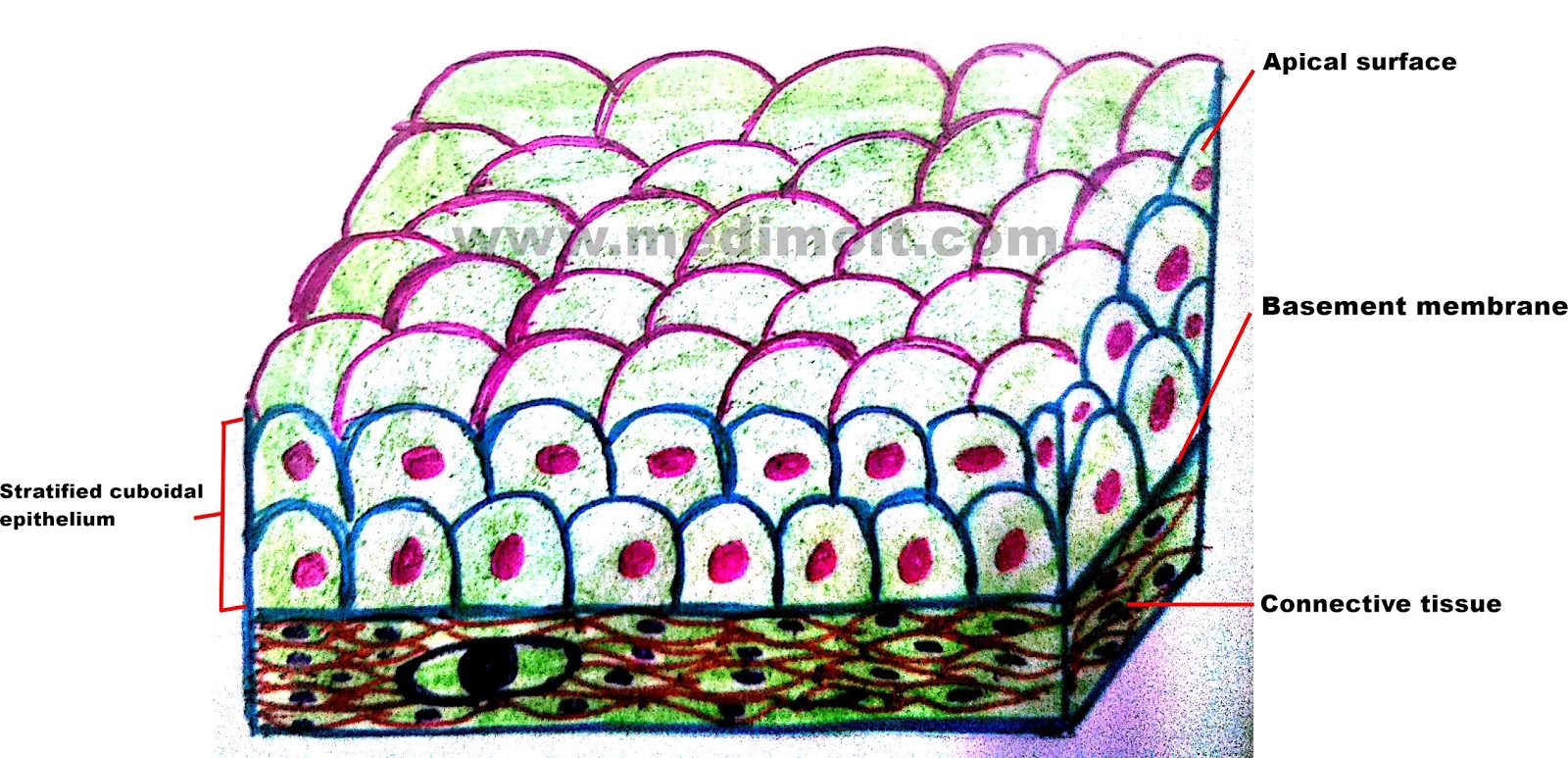
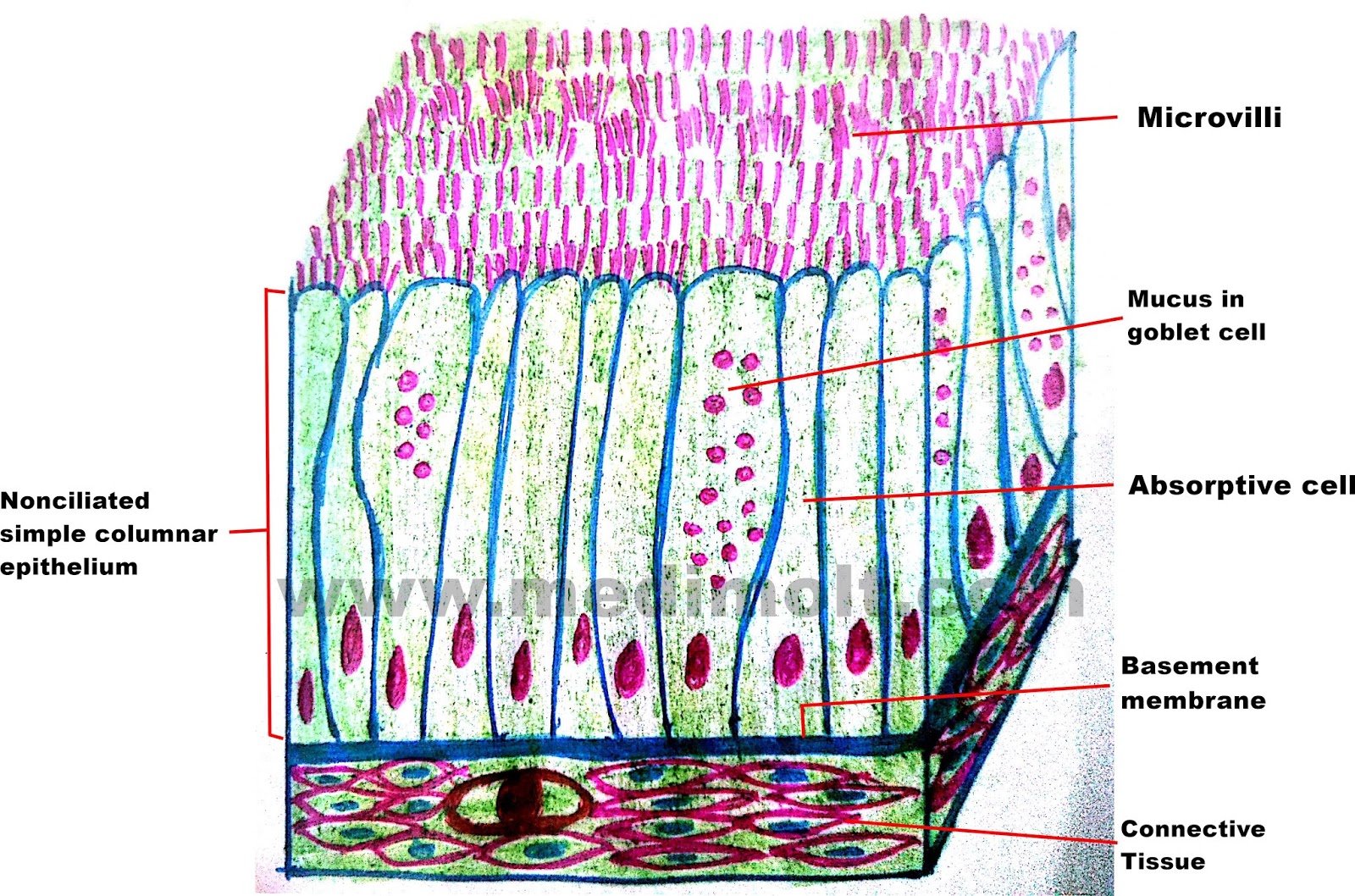
Mark 3370 module 5 quizzes (ch 13,14,9,10) 41 terms. The stratified squamous epithelium consists of several layers of cells, where the cells in the apical layer and several layers present deep to it are squamous, but the cells in deeper layers vary from cuboidal to columnar. Stratified cuboidal epithelium allows glands and organs to create a separation between the functioning cells of organ or gland and the vessels that feed it. Bodytomy provides a labeled diagram to help you understand the structure and simple columnar epithelium:
Tissue diagram stratified squamous epithelium.
Simple epithelium is one of the types of epithelium that is divided into simple columnar epithelium, simple squamous epithelium, and simple cuboidal epithelium. Bodytomy provides a labeled diagram to help you understand the structure and function of simple columnar. When the bladder is empty, the surface epithelial cells. Again, in the stratified epithelium, you will find more than one row of cells or nuclei.
The basal layer of the epithelium is attached to the basement membrane.
Here, the surface of the palatine tonsil covers the nonkeratinized stratified squamous epithelium. The oesophagus is an example of a stratified squamous non keratinising epithelium. Layer that is exposed to the outside of the cell. The cells in the apical layer and several layers deep to it are squamous while the cells in deeper layers vary from cuboidal to columnar.
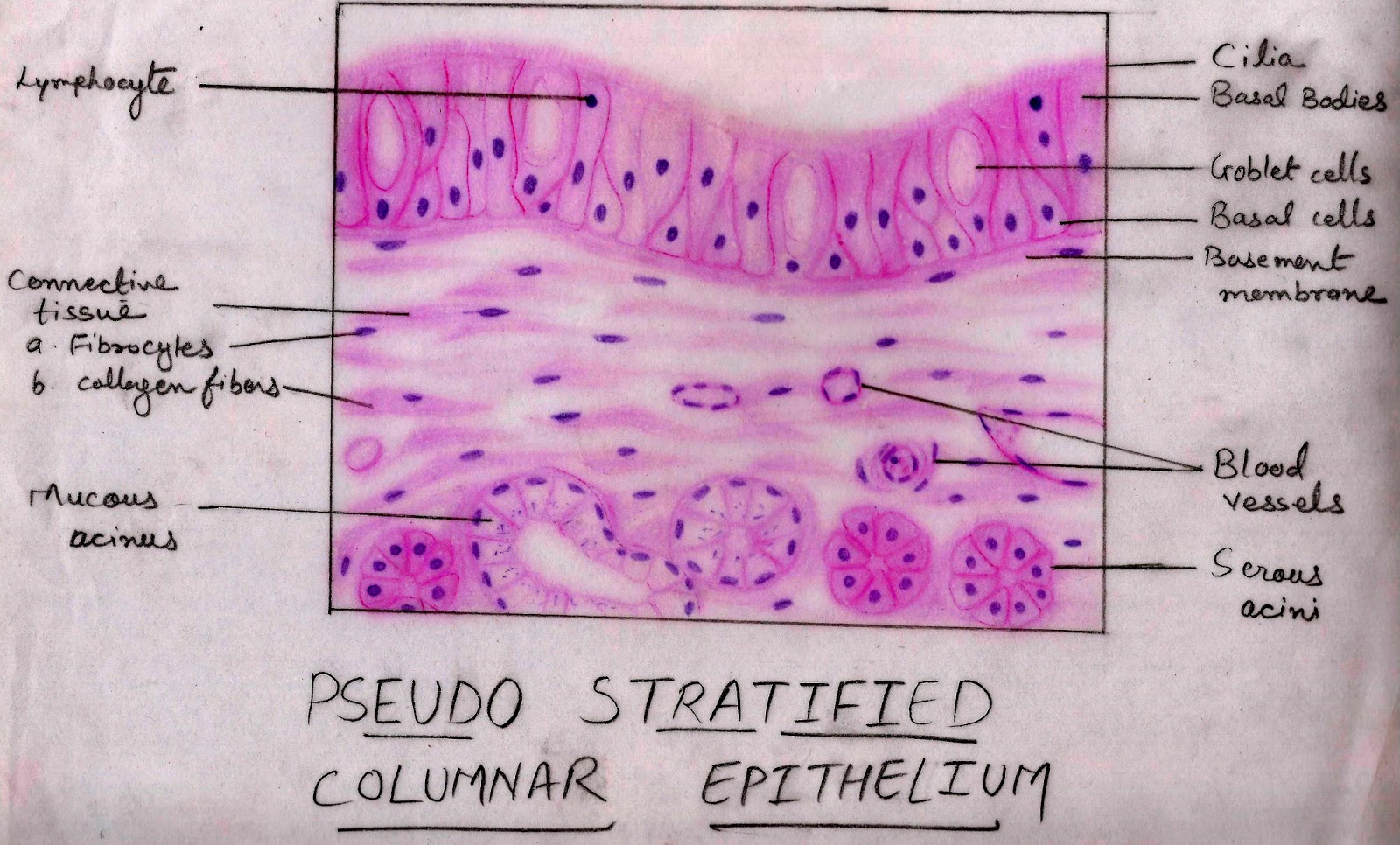
Pseudostratified columnar epithelium under a microscope this is not a true stratified epithelium but appears to be stratified.
They are specialised for secretion. Learn vocabulary, terms, and more with flashcards, games, and other study tools. It is present on almost every part of the human body, hence it has several important functions. The stratified columnar epithelium has multiple layers of cells in which the apical layer is made up of columnar cells while the deeper layer can be either cuboidal or columnar.
As in the case of other stratified epithelium, the cells in the deeper layers might be different than the layer on the top.
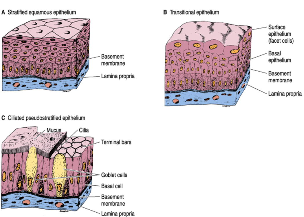
Start studying stratified cuboidal epithelium. Layer where cells are continually dividing by mitosis. Transitional epithelium is also called uroepithelium or urothelium (because it lines the urinary system), and it is a type of stratified epithelial tissue in which the surface cells change shape from being rounded to squamous in nature.transitional epithelium is located in the urinary system, especially the urinary bladder. Structure of stratified cuboidal epithelium.
Learn vocabulary, terms, and more with flashcards, games, and other study tools.
As the most important difference between the simple epithelium and the stratified epithelium is the number of the layer of cells, the functions of these layers also. Layers at the surface where keratin cements the debris of dead squamous cells, the non living keratin layer. The tonsillar surface form the deep grooves (known as the tonsillar crypts). So, two or three layers of crowded nuclei are seen in the pseudostratified columnar cells.
The layers of squamous epithelial cells beneath the cuboidal cells replace damaged cells as needed to maintain the epithelial lining.
Diagram of a stratified epithelium. The lower, deeper layers can be both cuboidal or columnar in shape. The glandular epithelium is made up of cuboidal or columnar cells. Start studying stratified columnar epithelium.
For example, it has roles in protection, absorption, secretion, and sensation.
Again, the diagram also shows the cells of the different layers of skin’s epidermis. A stratified squamous epithelium is a tissue formed from multiple layers of cells resting on a basement membrane, with the superficial layer(s) consisting of squamous cells. The complete viral life cycle is restricted to the differentiated cells of the epithelium. The modification of the cells on the apical surface is based on the location and function of.
But, in the longitudinal section of pseudostratified, the nuclei usually appear at a different level.
Stratified refers to how the epithelial tissue has layers. Its dominant presence also suggests that there are various. Stratified squamous diagram photo of endothelial cells.


:background_color(FFFFFF):format(jpeg)/images/article/en/stratified-epithelium/kxsJZMc6RS4j61ethfEOw_A4WBRjTb2LJJRtmn2wVxCw_stratified_squamous_epithelia02.png)


