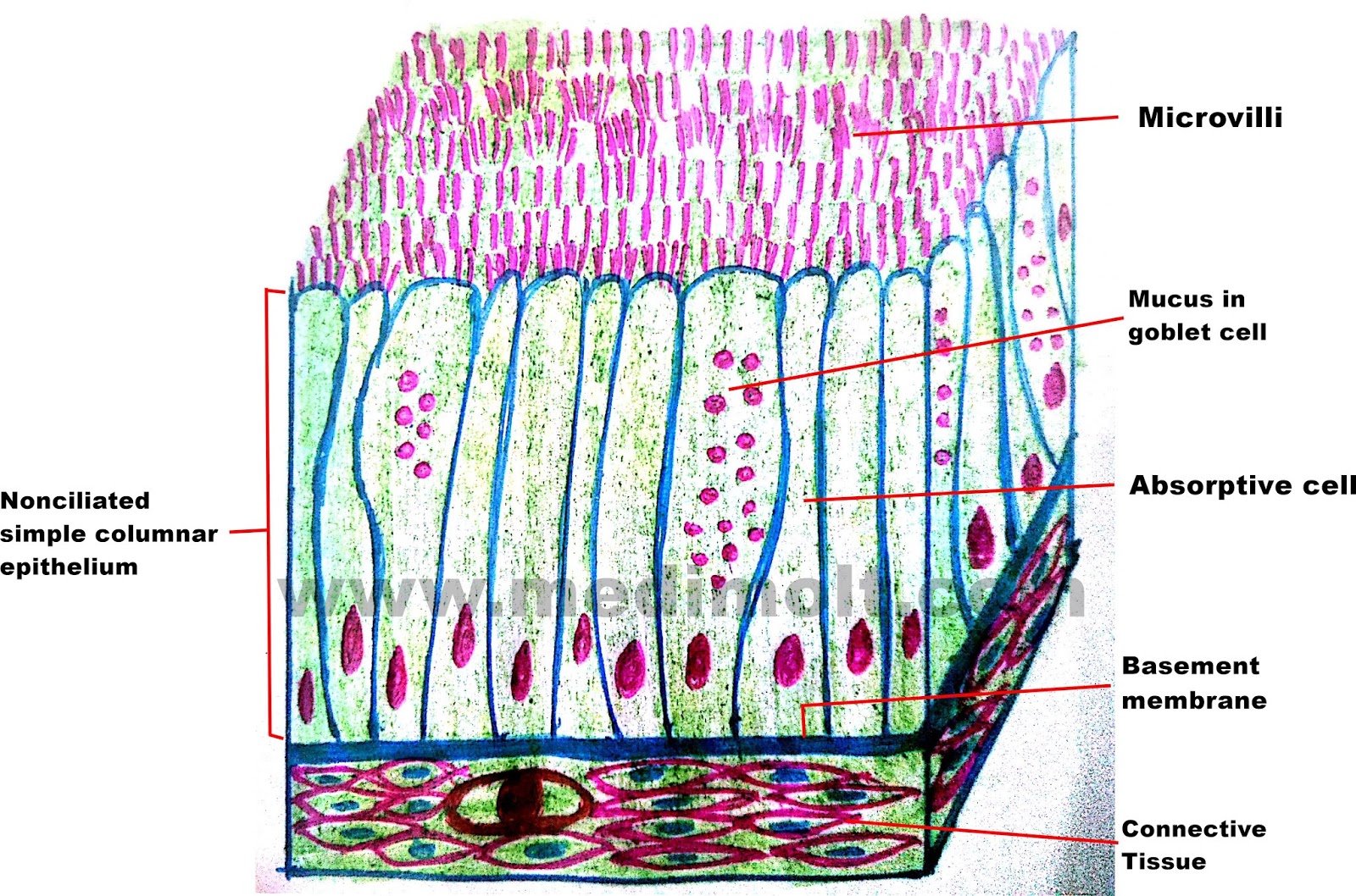
Epithelial tissue is classified based on the shape of the cells at the apical surface and the number of layers of cells. Cell sin the apical layer are flat; Appearance (falsely) of a stratified layer.
Schematic drawing (top) and actual image (bottom) of
» activity one — epithelium tissue observe the following tissues and sketch a drawing in the space provided below.
Made up of several layers of cells, continuously sloughed off and regenerated.
Simple squamous epithelium tissue stratified squamous epithelium tissue (cheek cell) (skin) simple cuboidal epithelium tissue simple columnar epithelium tissue Note the flattened nuclei and scant cytoplasm of the squamous epithelium. The lower layer is columnar and metabolically active: The lower, deeper layers can be both cuboidal or columnar in shape.
In the cuboidal epithelium, notice that the cells are about as tall as they are wide, the nuclei are.
The apical layer and several layers deep to it are partially d…. This tissue consists of multiple layers of cube shaped cells. Learn vocabulary, terms, and more with flashcards, games, and other study tools. Those of the deep layers v….
This type is relatively rare, occurring specifically in the lining of excretory ducts, such as salivary and sweat glands.
From your observations, do you think stratified squamous epithelium is the; Reset pseudostratified simple columnar epithelium simple squamous epithelium stratified cuboidal epithelium simple epithelia columnar epithelium stratified squamous stratified squamous keratinized epithelium simple cuboidal epithelium stratified columnar. The older layer of cells is pushed upwards and becomes flat. Like all epithelial tissue, stratified cuboidal epithelium is avascular, which means that it lacks blood vessels.
Summary of epithelial tissues art a rag the appropriate labels to their respective targets.
Start studying epithelial tissue images and drawings. Nuclei within cells whose height and width are similar. The multiple layers of stratified squamous moist epithelium provide protection against friction and trauma to organs within the body. Two or more layers of cells;
Transitional epithelium, often called urinary epithelium, lines the urinary tract and, therefore, must undergo transitions.
Cells) and simple cuboidal epithelium lines the tubules (tips of blue arrows). Mammary glands, sweat gland and salivary glands Transitional epithelium, with its multiple layers, protects underlying tissues from urine that is stored in the urinary bladder. Structure of stratified cuboidal epithelium.
Large ducts in most exocrine glands structure:
Thus, this epithelium varies between stratified cuboidal and stratified squamous. Types of epithelial tissues (10. Stratified cuboidal epithelial tissues are more rare comparing to other epithelial types. The lining of the mouth and vagina:
Epithelium, noncornified typical of mucosal surfaces, embryonic germ layers including endoderm, protection serves stratified squamous, embryonic germ layers including mesoderm, multiple several cell shapes cuboidal, transitional epithelium e.g.
Cells at apical surface are cuboidal basement membrane apical surface basal surface function: Bladder lining, simple cuboidal form glands,. The major purpose of this type of epithelium is to secrete and protect the body. They can be found in the eye's conjunctiva coating.
Lines mouth and esophagus, does not contain keratin in the api….
Epithelial tissue classification table classification function location identification sketch class notes simple squamous epithelium simple cuboidal epithelium simple columnar epithelium pseudostratified ciliated columnar epithelium stratified squamous epithelium without keratin stratified squamous epithelium with keratin stratified cuboidal,. Up to 24% cash back a keratinized type of stratified squamous epithelium is found in the epidermis of the skin,. The basal layer of the epithelium is attached to the basement membrane. Use it in your personal projects or share it as a cool sticker on whatsapp, tik tok, instagram, facebook messenger, wechat, twitter or in other messaging apps.
Transitional epithelium also transitions from looking like stratified cuboidal when the bladder is empty to looking like stratified
Download now for free this stratified cuboidal epithelium text transparent png image with no background. They mostly line the sweat gland excretory ducts, which are huge ducts. Stratified cuboidal epithelial tissue from salivary gland (tm: This picture was taken from salivary gland and the duct showed that the inner most layer, or right around the lumen, contains cuboidal cells but the rest of the layers may or may not be cuboidal in shape.
The cells are tightly packed to ensure no gap is present in two cells.
This concept map, created with ihmc cmaptools, has information related to: There is also a large, round central nucleus in each cell. The major purpose of this type of epithelium is to protect the body. Stratified squamous, nonkeratinized epithelium this image demonstrates the transition of epithelial cells from cuboidal at the basement membrane to squamous cells at the surface.
Compare your drawing of simple cuboidal epithelium from the previous activity to your sketch of stratified squamous epithelium in this activity.
In general, epithelial tissue is any group of cells lining a body cavity or body surface. Stratified cuboidal epithelium is quite thin, consisting of two or three layers of cuboidal cells. Its main function is structural reinforcement, since it is not significantly involved in absorption or secretion. Sketch a few cells of each type of epithelium you observed.
This is a fairly rare type of epithelium in which the cells in….


