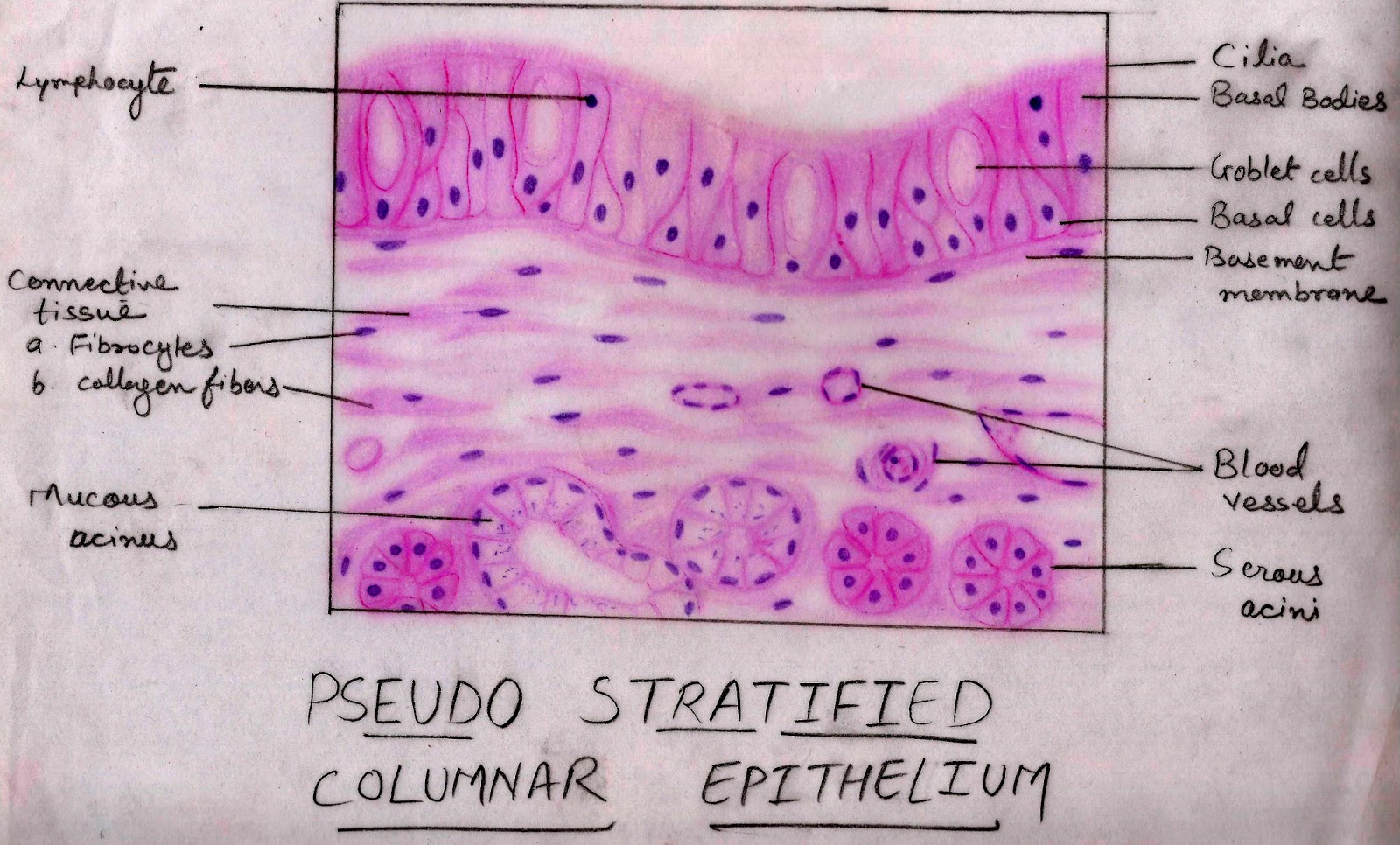
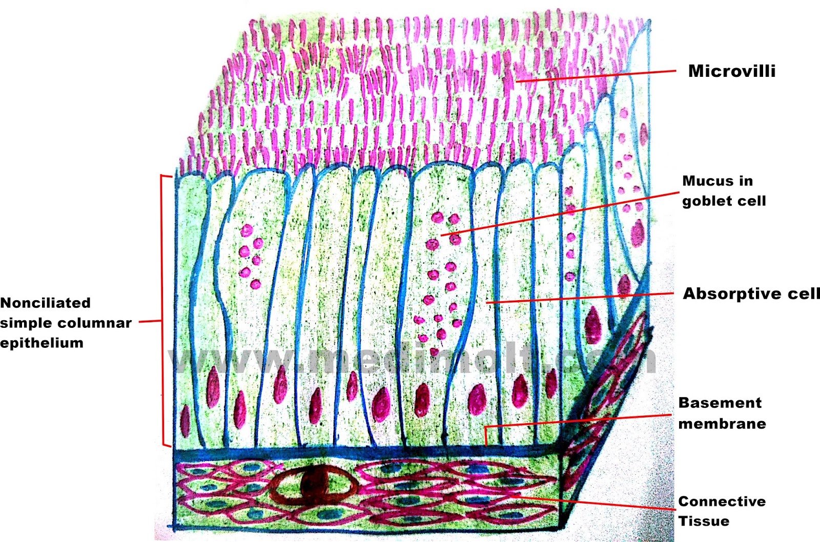
Remember that these slides show slices from an organ, and that organs are made up of several different types of tissues. The modification of the cells on the apical surface is based on the location and function of. As in the case of other stratified epithelium, the cells in the deeper layers might be different than the layer on the top.
Pin by Maria Kozlova on Biology&medicine Tissue types
The stratified cuboidal epithelium is composed of multiple layers of cells, in which the topmost layer is composed of cuboidal cells, while the lower layer could be columnar or cuboidal.
(layers), and are called stratified.
Label the parts of the stratified cuboidal epithelium. Identify the structure labeled 3. Experts are tested by chegg as specialists in their subject area. Read more about stratified squamous epithelium, esophagus (40x) 1 comment;
The nuclei of the cuboidal epithelial cells are round and generally located in the center of the cell.
Synovial membrane of synovial joint. This type is relatively rare, occurring specifically in the lining of excretory ducts, such as salivary and sweat glands. Simple cuboidal epithelium histology slide #3. Draw the significant features 2.
Structure of stratified cuboidal epithelium.
A labeled diagram and functions epithelium is a tissue that lines the internal surface of the body, as well as the internal organs. Topmost layer of skin epidermis in frogs, fishes is made up of living cuboidal cells. Simple epithelium is one of the types of epithelium that is divided into simple columnar epithelium, simple squamous epithelium, and simple cuboidal epithelium. Label the letters below simple cuboidal epithelium simple squamous epithelium stratified cuboidal epithelium.
Stratified cuboidal epithelium describes an epithelial tissue with two aspects.
Only the most superficial layer is made up of cuboidal cells, and the other layers can be cells of other types. Correctly identify this tissue type and then label the features of the tissue. Definition of stratified cuboidal epithelium. The basal layer of the epithelium is attached to the basement membrane.
They protect areas such as ducts of sweat glands, mammary glands, and salivary glands.
Simple squamous histology slide #2. (3) squamous, thin, flat cells that look like the scales of a fish from a superior view. We review their content and use your feedback to keep the quality high. In the stratified squamous epithelium labeled diagram, i tried to show you the columnar cells, elongated nucleus, and basement membrane.
Stratified cuboidal epithelia is a rare type of epithelial tissue composed of cuboidally shaped cells arranged in multiple layers.
Stratified cuboidal epithelium is a type of epithelial tissue found mainly in glands, which specialize in selective absorption and secretion by the gland into blood or lymph vessels. Has cells of approximately equal height and width with spherical nuclei that are centered in the cell. Label the structures listed 3. Name the locations where the tissue is found a.
There are three general shapes of epithelial cells:
100% (15 ratings) the parts of str. Simple columnar epithelium histology slide #4. The other layers may contain cells that are cuboidal and/or columnar, but the classification of the epithelium is based only on the shape of the outermost layer of cells. Is generally found lining small tubes and ducts with absorptive, excretory and/or secretory functions.
Secretion from the closely associated glands lubricates the surface of the nonkeratinized epithelium.
Identify the epithelial tissue labeled 1. Basement connective epithelial tissue lumen free surface nucleus. Total magnification 400x, objective 40x. In general, epithelial tissue is any group of cells lining a body cavity or body surface.
The lower, deeper layers can be both cuboidal or columnar in shape.
Simple cuboidal epithelium lines small ducts as seen here. The cells are tightly packed to ensure no gap is present in two cells. The stratified columnar epithelium has multiple layers of cells in which the apical layer is made up of columnar cells while the deeper layer can be either cuboidal or columnar. Because all of the cells are directly attached to the basement membrane (thin blue line)
Identify the structure labeled 2.
Ciliated epithelium is a thin tissue that. Epithelium stratified cuboidal stratified squamous epithelium epithelium connective tissue squamous cells simple cuboidal epithelium cuboidal cells columnar cells name this tissue type: There is also a large, round central nucleus in each cell. Like all epithelial tissue, stratified cuboidal epithelium is avascular, which means that it lacks blood vessels.
Its main function is structural reinforcement, since it is not significantly involved in absorption or secretion.
Epithelial tissues identify the following epithelial tissues under the microscope. From the epithelial tissue, i should learn and identify the following histology slides with their identification points. Stratified squamous epithelium (nonkeratinized) identify the epithelial tissue. Stratified squamous epithelia consist of multiple layers of cells with the outer most layer being squamous.
Labeled diagram simple cuboidal epithelial cells are shaped like cubes, and the nucleus of each cell is large and located close to the center of the cell.
Stratified cuboidal epithelium is quite thin, consisting of two or three layers of cuboidal cells.


