Structural organization human anatomy and physiology organic chemistry study basement membrane. Anatomynote.com found stratified cuboidal epithelium from plenty of anatomical pictures on. Transitional epithelium is also called uroepithelium or urothelium (because it lines the urinary system), and it is a type of stratified epithelial tissue in which the surface cells change shape from being rounded to squamous in nature.transitional epithelium is located in the urinary system, especially the urinary bladder.
What is Epithelial Tissue Different Types of Structure
There is also a large, round central nucleus in each cell.
100% (15 ratings) the parts of str.
The basal layer of the epithelium is attached to the basement membrane. This epithelium protects against physical and chemical damage. Stratified cuboidal epithelium describes an epithelial tissue with. Start studying stratified cuboidal epithelium.
Function (stratified cuboidal epithelium) protection.
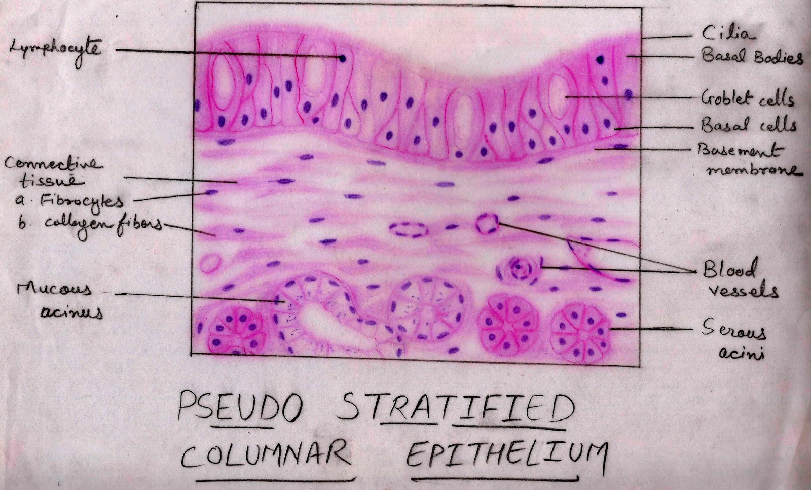
Stratified cuboidal epithelial tissues are more rare comparing to other epithelial types. Basement connective epithelial tissue lumen free surface nucleus. Stratified cuboidal epithelium study guide by emily_rossetto includes 6 questions covering vocabulary, terms and more. Pseudostratified columnar of paranasal sinuses paranasal sinuses are the bilateral closed cavities in the frontal, maxillary,.
Anatomy and physiology questions and answers.
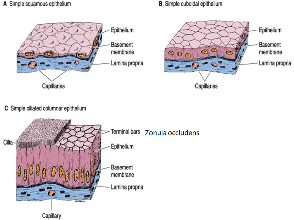
Location (stratified cuboidal epithelium) largest ducts of sweat glands, mammary glands, and salivary glands. They have equal dimensions on all sides. Simple epithelium is one of the types of epithelium that is divided into simple columnar epithelium, simple squamous epithelium, and simple cuboidal epithelium. In general, epithelial tissue is any group of cells lining a body cavity or body surface.
Of unknown organism, from personal collection.
This set is often in folders with. We review their content and use your feedback to keep the quality high. Keratin is a tough fibrous intracellular protein that helps protect skin and underlying tissues from heat microbes and chemicals. Calculus for the life sciences 1st.
Connective tissue basal lamina stratified cuboidal epithelium nucleus lumen of duct labels to the appropriate location in the figure.
It is mostly responsible for protection and mucous secretion. Stratified cuboidal cells might be square, round or hexagonal in shape. Labeled diagram simple cuboidal epithelial cells are shaped like cubes, and the nucleus of each cell is large and located close to the center of the cell. The cells are tightly packed to ensure no gap is present in two cells.
Collagen fibers (dense irregular) collagen fibers (dense regular) fibroblast.
Stratified cuboidal epithelium is a type of epithelial tissue found mainly in glands, which specialize in selective absorption and secretion by the gland into blood or lymph vessels. A labeled diagram and functions epithelium is a tissue that lines the internal surface of the body, as well as the internal organs. The lower, deeper layers can be both cuboidal or columnar in shape. Ciliated epithelium is a thin tissue that.
This picture was taken from salivary gland and the duct showed that the inner most layer, or right around the lumen, contains cuboidal cells but the rest of the layers may or.
We are pleased to provide you with the picture named stratified cuboidal epithelium.we hope this picture stratified cuboidal epithelium can help you study and research. Learn vocabulary, terms, and more with flashcards, games, and other study tools. Label the parts of the stratified cuboidal epithelium. Tissue diagram stratified squamous epithelium.
For more anatomy content please follow us and visit our website:
In the stratified squamous epithelium labeled diagram, i tried to show you the columnar cells, elongated nucleus, and basement membrane. Structure of stratified cuboidal epithelium. Experts are tested by chegg as specialists in their subject area. Part a drag the labels onto the diagram to identify structural features of epithelium.
A stratified epithelium consists of multiple stacked layers of cells.
Transitional epithelium location and characteristics Form the inner lining of blood vessels, ducts and body cavities, and the interior of the respiratory, digestive, urinary and reproductive systems. Download scientific diagram | stratified cuboidal epithelium composed of several layers of cuboidal cells and lacking ciliated cells. Constitute the secretory portion of glands.
It is located in the conjunctiva inside the eyelids and areas of tissue transition.
Stratified squamous epithelium is the most common type of stratified epithelium in the human body. When the bladder is empty, the surface epithelial cells of. The stratified epithelium is named by the shape of the most apical layer of cells, closest to the free space.