Look for these sweat glands well beneath the surfacing epithelium. As protection against desiccation, it undergoes a process known as cornification or keratinization. The cells are tightly packed to ensure no gap is present in two cells.
simple cuboidal epithelium Nursing study, Stevenson
By convention, a stratified epithelium is described according to the shape of cells on its free surface.
This epithelium is found at the surface of the skin and is known as the epidermis.
This esophagus has an example of a large duct with a stratified cuboidal epithelium. But because each cell rests on the. This epithelium covers internal body surfaces which are exposed to some degree of physical trauma. Stratified cuboidal epithelium is found in the ducts of sweat glands.
There are three principal shapes of epithelial cell:
Simple cuboidal epithelium histology slide #3. Epithelial cells make up primary tissues throughout the body. This histology test bank is also useful for the histology questions on the usmle (usmle step 1). The basal layer of the epithelium is attached to the basement membrane.
Stratified cuboidal epithelium is a type of epithelial tissue found mainly in glands, which specialize in selective absorption and secretion by the gland into blood or lymph vessels.
Is pretty rare, found around large excretory ducts of the pancreas, salivary glands, and sweat glands. For each histology question, pick the one best answer. Click here for answers and detailed explanations. The cells of the basal layer are cuboidal, while the cells at the surface are columnar, thus the name stratified columnar.
Thus, this epithelium varies between stratified cuboidal and stratified squamous.
Provides a stronger lining than simple epithelium. It is most often found in large ducts from exocrine glands. Simple columnar epithelium histology slide #4. Pseudostratified ciliated columnar epithelium slide #7.
Slide 106 (plantar skin, h&e) view virtual slide slide 112 (plantar skin, h&e view virtual slide.
Learn vocabulary, terms, and more with flashcards, games, and other study tools. A basement membrane separates the epithelium from underlying connective tissue. This epithelium shows a basal layer of. Number of cell layers (single or compound)
Smaller ducts with a stratified cuboidal epithelium are also present.
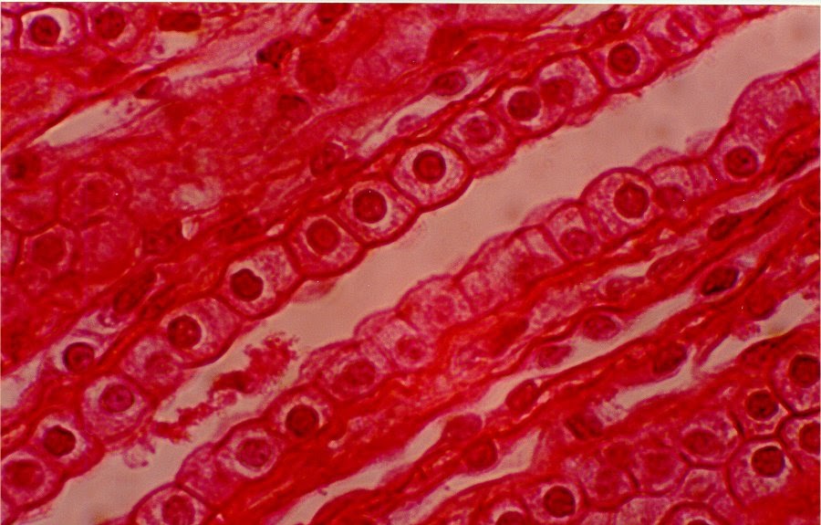
Epithelium forms continuous sheets of cells that line internal surfaces and cover the external surface of the body. Slide 43 skin, sole of foot. Epithelial cells form from ectoderm, mesoderm, and endoderm, which explains. Stratified cuboidal of the sweat gland ducts, 150x.
This epithelium shows a basal layer of cuboidal cells, then several layers of polygonal cells that become progressively more flat, until they become squamous at the luminal surface.
Click again to see term 👆. Epithelium is classified based on three criteria: Stratified squamous, nonkeratinized epithelium this image demonstrates the transition of epithelial cells from cuboidal at the basement membrane to squamous cells at the surface. It acts as a selective barrier that protects tissues.
In general, epithelial tissue is any group of cells lining a body cavity or body surface.
Stratified cuboidal epithelium describes an epithelial tissue with two aspects. Such transitions occur in the ducts of larger glands. These ducts range from simple cuboidal, simple columnar, to stratified cuboidal epithelia. Name the epithelium sweat gland ducts, large exocrine ducts.
Name the epithelium (primary one in the center of image).
Stratified cuboidal epithelium has a limited distribution. It most often has only two layers of cuboidal cells. Usually has 2 or 3 layers of cells. Pseudo stratified signifies that two or more rows of nuclei give the (false) appearance of a stratified epithelium.
Typically, it has only two layers of cuboidal cells.
Also observe some stratified cuboidal epithelium lining the larger ducts found in this section (typically lying within connective tissue septa). These can be arranged in a single layer of cells as simple epithelium, either squamous, columnar, or cuboidal, or in layers of two or more cells deep as stratified (layered), or compound, either squamous, columnar or cuboidal. Structure of stratified cuboidal epithelium. Is not involved in absorption.
Regardless of whether the surface cells are squamous, cuboidal, or columnar, the underlying cells are usually cuboidal.
The lower, deeper layers can be both cuboidal or columnar in shape. The multiple layers of stratified squamous moist epithelium provide protection against friction and trauma to organs within the body. Stratified cuboidal epithelium has a limited distribution but is prominent in ducts of exocrine glands. In some tissues, a layer of.
This image shows a section through a sweat gland and its duct, lined by a stratified cuboidal epithelium.
Stratified cuboidal eptiehlium slide #6. There are many arrangements of epithelial cells such as squamous, cuboidal, and columnar that organize as simple, stratified, pseudostratified, and transitional.