The lower, deeper layers can be both cuboidal or columnar in shape. Male urethra and lobar ducts of salivary glands: The cells are tightly packed to ensure no gap is present in two cells.
Stratified Cuboidal Epithelium Function slidedocnow
Collagen fibers (dense irregular) collagen fibers (dense regular) fibroblast.
Generally two layers of cubelike cells.
Content may be subject to copyright. The nuclei are rounded and lie in the centre of the cells. At the tip of the black arrow is stratified cuboidal epithelium. The given figure is of simple cuboidal epithelium.
Secretory duct of sweat glands, mammary glands and sebaceous gland, pancreas, salivary glands;
Outermost layer of cells are cube like, cells are nucleated & living. Anatomy and physiology questions and answers. A typical example of stratified squamous keratinized epithelium is the epidermis. The modification of the cells on the apical surface is based on the location and function of.
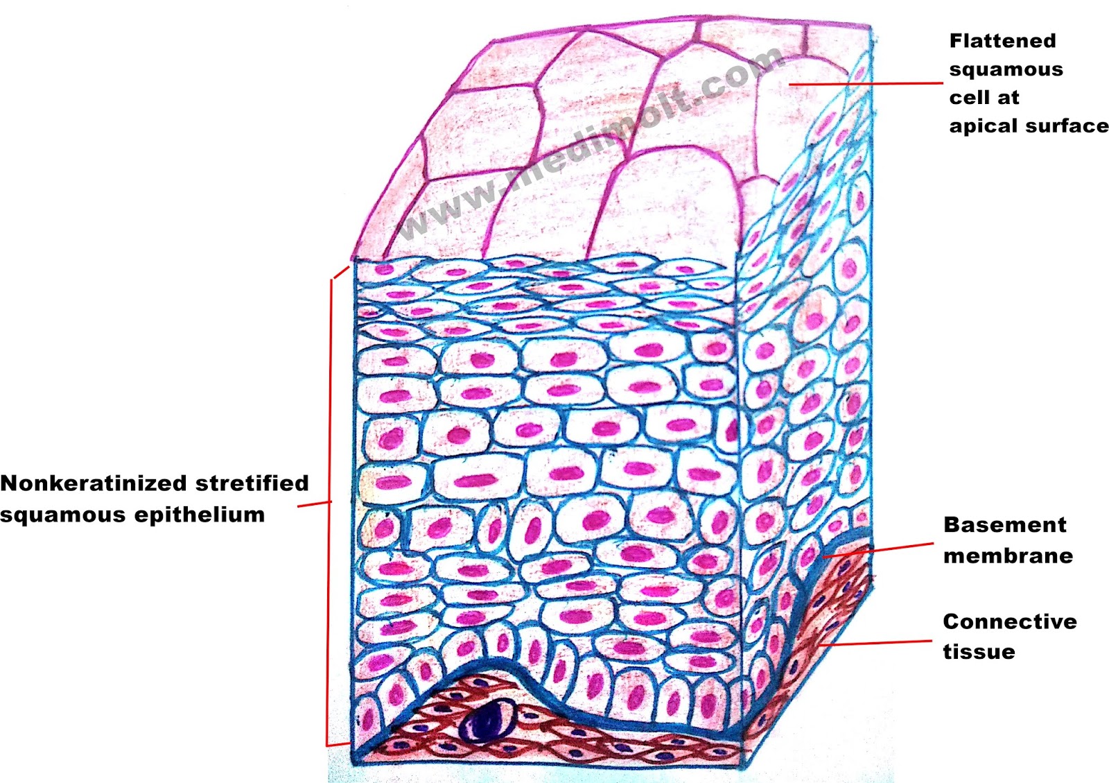
Transitional epithelium is also called uroepithelium or urothelium (because it lines the urinary system), and it is a type of stratified epithelial tissue in which the surface cells change shape from being rounded to squamous in nature.transitional epithelium is located in the urinary system, especially the urinary bladder.
Stratified cuboidal epithelium (h & e). The cuboidal epithelium is present in the small salivary and pancreatic ducts, thyroid follicles, parts of membranous. They have equal dimensions on all sides. Mammary glands, sweat gland and salivary glands:
Labeled diagram simple cuboidal epithelial cells are shaped like cubes, and the nucleus of each cell is large and located close to the center of the cell.
Anatomy and physiology questions and answers. There are three types of epithelial cells which differ in their shape and function. Explore smile with your eyes' photos on flickr. When you sweat, this layer of stratified cuboidal epithelium allows various salt ions and water to slip into the vessel.
The upper layer is cuboid and other layers may be cuboidal or other types:
The cells in the apical layer and several layers deep to it are squamous while the cells in deeper layers vary from cuboidal to columnar. Protection of ducts of various glands: Tissue diagram stratified squamous epithelium. There is also a large, round central nucleus in each cell.
In this image, you will find basement membrane, cuboidal epithelial cells, duct lumen in stratified cuboidal epithelium.
Connective tissue basal lamina stratified cuboidal epithelium nucleus lumen of duct labels to the appropriate location in the figure. This empty space is a small vessel which leads out of the skin. Largest ducts of sweat glands, mammary gland, and salivary glands. Download scientific diagram | stratified cuboidal epithelium composed of several layers of cuboidal cells and lacking ciliated cells.
The oesophagus is an example of a stratified squamous non keratinising epithelium.
Part a drag the labels onto the diagram to identify structural features of epithelium. There is a layer of columnar cells present on squamous, columnar or cuboidal epithelial cells The cells are cubical and more or less squarish in shape. H a) a b) b c) c d) d e) e f) f g) g h) h 6) in the photomicrograph shown below, which layer will contain melanocyte projections, keratinocytes and intraepidermal macrophages.
This type is relatively rare, occurring specifically in the lining of excretory ducts, such as salivary and sweat glands.
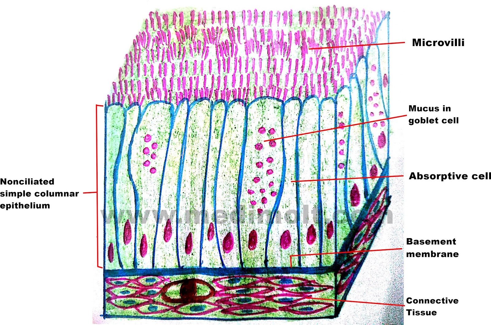
Simple epithelium is one of the types of epithelium that is divided into simple columnar epithelium, simple squamous epithelium, and simple cuboidal epithelium. When the bladder is empty, the surface epithelial cells of. A labeled diagram and functions epithelium is a tissue that lines the internal surface of the body, as well as the internal organs. The basal layer of the epithelium is attached to the basement membrane.
Structure of stratified cuboidal epithelium.
Inner most layer is cuboidal; Stratified squamous epithelium diagram and structure it has two or more layers of cells. In surface view, the cells appear polygonal. The stratified columnar epithelium has multiple layers of cells in which the apical layer is made up of columnar cells while the deeper layer can be either cuboidal or columnar.
Outermost layer is composed of pillar shaped cells, cells are nucleated.
As in the case of other stratified epithelium, the cells in the deeper layers might be different than the layer on the top. 5) in the diagram of skin shown below, which structures are composed of stratified cuboidal tissue? Stratified cuboidal cells might be square, round or hexagonal in shape.