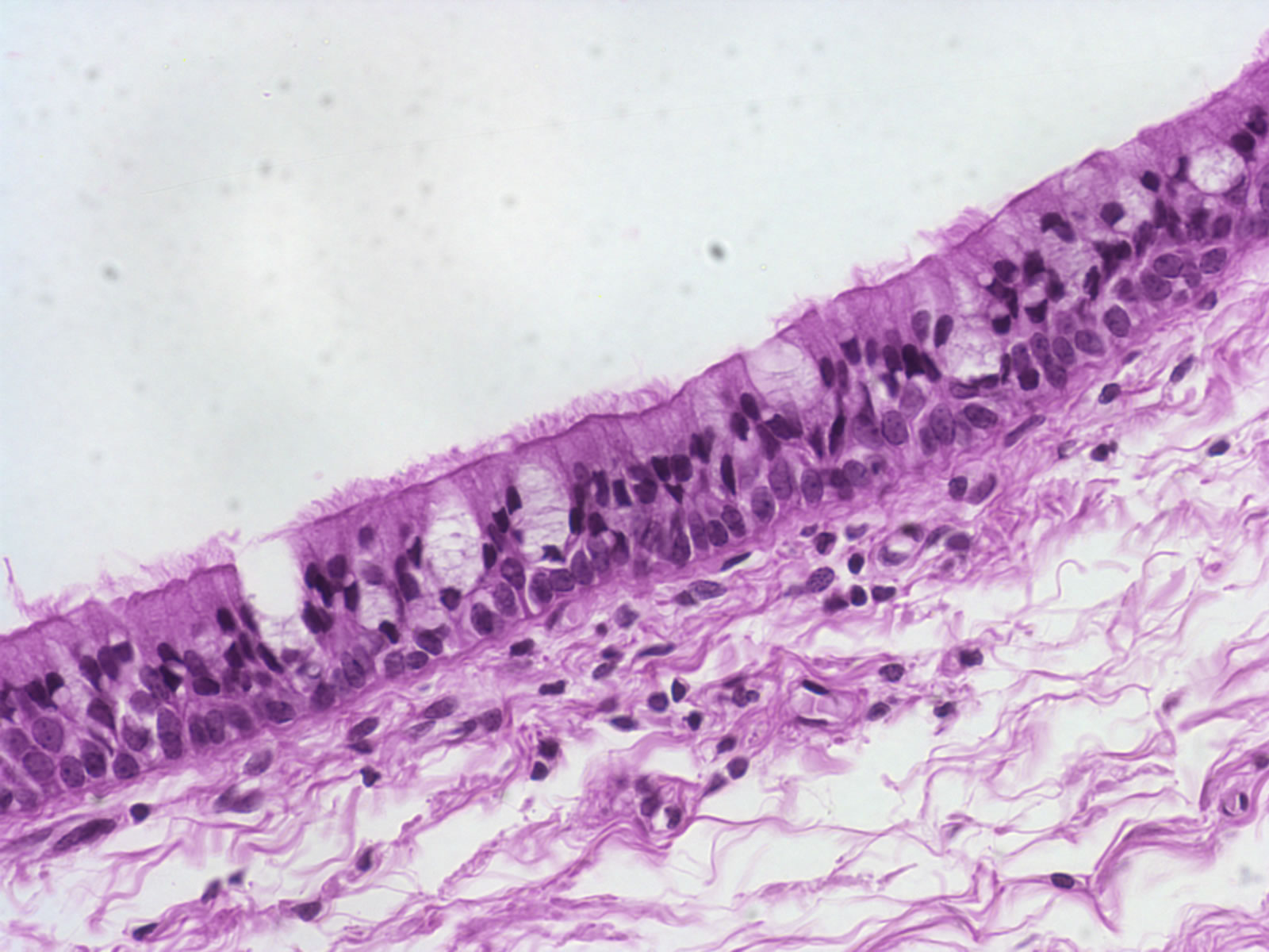
While the efferent ducts are more scalloped in appearance. The modification of the cells on the apical surface is based on the location and function of. In the deep layer of stratified squamous epithelium, you will find the columnar epithelium that rests on the basement membrane.
Microscopic Image Of Colored Stratified Squamous
Cytoplasm (simple columnar epithelium) what is the green.
But, in the longitudinal section of pseudostratified, the nuclei usually appear at a different level.
Stratified squamous epithelium (keratinizing and nonkeratinizing): Tissues to identify under the microscope. Under a microscope, epithelial cells are readily distinguished by the following features: Stratified ciliated columnar epithelium prepared microscope slide.
In the case of stratified squamous epithelium cells in the layers below may differ from the epithelium on top.
Observing the pseudostratified columnar cells under the light microscope will find more than. Unlike the epithelium of the skin, a pseudostratified ciliated columnar epithelium appears to have multiple layers, but is actually only comprised of a single sheet of cells. O the cell membrane is usually not clearly seen under the light microscope. In pseudostratified epithelium, nuclei of neighboring cells appear at different levels rather than clustered in the basal end.
As in the case of other stratified epithelium, the cells in the deeper layers might be different than the layer on the top.
Simple secretory columnar epithelium lines the stomach and uterine schematron.org simple columnar epithelium that lines the intestine also contains a few goblet cells. Epithelia are tissues consisting of closely apposed cells without intervening intercellular substances. Basal cells of stratified squamous epithelium. Epithelial tissue covers or lines body surfaces as well as serving to absorb, filtrate, protect, and secrete various substances.
So, two or three layers of crowded nuclei are seen in the pseudostratified columnar cells.
Make sure this fits by entering your model number. The positioning of the nuclei within the individual columnar cells causes this illusion. 7 m h&e microscope slide. O topmost = apical part = luminal part of the.
Therefore, the shape of a cell is identified by the appearance of its nucleus.
Just underneath it you can see a layer of connective tissue (ct). Light micrograph of a section through stratified columnar epithelium (across upper centre). 7 µm h&e microscope slide | carolina.com. The epididymis has a smooth, even epithelium lining its lumen;
Learn vocabulary, terms, and more with flashcards, games, and other study tools.
The epithelium was examined under the light microscope at 4 points in each region and the type of epithelium was classified. Plasma membrane of adipose cell. The stratified columnar epithelium has multiple layers of cells in which the apical layer is made up of columnar cells while the deeper layer can be either cuboidal or columnar. Human stratified columnar epithelium, sec.
Both are examples of pseudostratified columnar epithelium.
Again, in the stratified epithelium, you will find more than one row of cells or nuclei. The cells that comprise the epithelial membranes are variously shaped and are named accordingly. Section of frog esophagus or oral mucosa. A labeled diagram and functions epithelium is a tissue that lines the internal surface of the body, as well as the internal organs.
These structures, which are easily identifiable with the help of a microscope, are found at various levels, creating.
As well as providing a protective barrier, it also has secretory functions. As implied by their moniker, columnar epithelial cells are taller than they are wide, appearing like numerous miniature pillars. A 10% discount applies if you order more than 10 of this item and 15% discount applies if you order more than 25 of this item. It is found in parts of the eye, pharynx, reproductive and urinary tracts, and salivary glands.
Squamous epithelial cells under microscope view.
Stratified columnar epithelium is often confused with pseudostratified columnar epithelial tissue, as they can look very similar under a microscope. Our results indicate that the human female urethra is lined by stratified squamous, pseudostratified columnar and, occasionally, transitional epithelium. Identify these cells of this tissue. Especially note the stereocilia (long microvilli) of the epididymis.
Pseudostratified columnar epithelium is a type of epithelium that appears to be stratified but instead consists of a single layer of irregularly shaped and differently sized columnar cells.
It would be more obvious under the microscope, and will be very easy to see on the next image. The cells in pseudostratified columnar epithelial tissue mimic stacked layers of columnar cells, but if one looks closely, the bottom of each cell connects with the basement membrane, so there is actually only one layer. This is a rare type of epithelial tissue (tissues that line the cavities and surfaces of the body). The tissue is classified by the number of cell layers it has (simple=1 cell layer, stratified = more than 1 cell layer) and the shape of the cells (squamous=flat,.
The stratified cuboidal epithelium is composed of multiple layers of cells, in which the topmost layer is composed of cuboidal cells, while the lower layer could be columnar or cuboidal.


