The tissues that comprise a single layer of cells are called simple, and the ones that have more than one layer are referred to as stratified. The fetal epithelial lining of the esophagus is a complex entity. The pseudostratified columnar epithelium comprises a single layer of cells but seems to be multilayered.
Mammalian Histology Epithelial Tissues Berkshire
Simple columnar or pseudostratified columnar tissue / organ:
They are also classified on the basis of the number of layers of cells.
This is a special type of simple epithelium called pseudostratified epithelium as it resembles stratified epithelium due to the positioning of the cellular nuclei, but is comprised of only a single layer of cells. Simple epithelium is one of the types of epithelium that is divided into simple columnar epithelium, simple squamous epithelium, and simple cuboidal epithelium. Pseudostratified epithelium is most commonly found in the form of columnar shaped epithelium, but can also. Simple columnar epithelium features label the features of simple.
It is located in the conjunctiva inside the eyelids and areas of tissue transition.
Pseudostratified columnar epithelium under a microscope with a labeled diagram. In the case of stratified squamous epithelium cells in the layers below may differ from the epithelium on top. The stratified columnar epithelium has multiple layers of cells in which the apical layer is made up of columnar cells while the deeper layer can be either cuboidal or columnar. Stratified columnar epithelium is a type of epithelial tissue composed of two or more layers of columnar epithelial cells.epithelial tissue is one of the body’s primary tissues and lines the surfaces and insides of structures in many parts of the body.
It is named for the shape of the cells on the surface of the tissue.
_____ check your understanding 1. Transitional epithelium location and characteristics Nucleus of a squamous cell, keratin layer locations: The cells in this tissue are not all squamous (flat).
Learn vocabulary, terms, and more with flashcards, games, and other study tools.
The arrow indicates one of these squamous cells. 100% (7 ratings) the apical cells of stratified columnar epithelium are columnar in sha. As in the case of other stratified epithelium, the cells in the deeper layers might be different than the layer on the top. Intermediate fetal epithelia include a pseudostratified ciliated epithelium that dominates the second trimester esophagus.
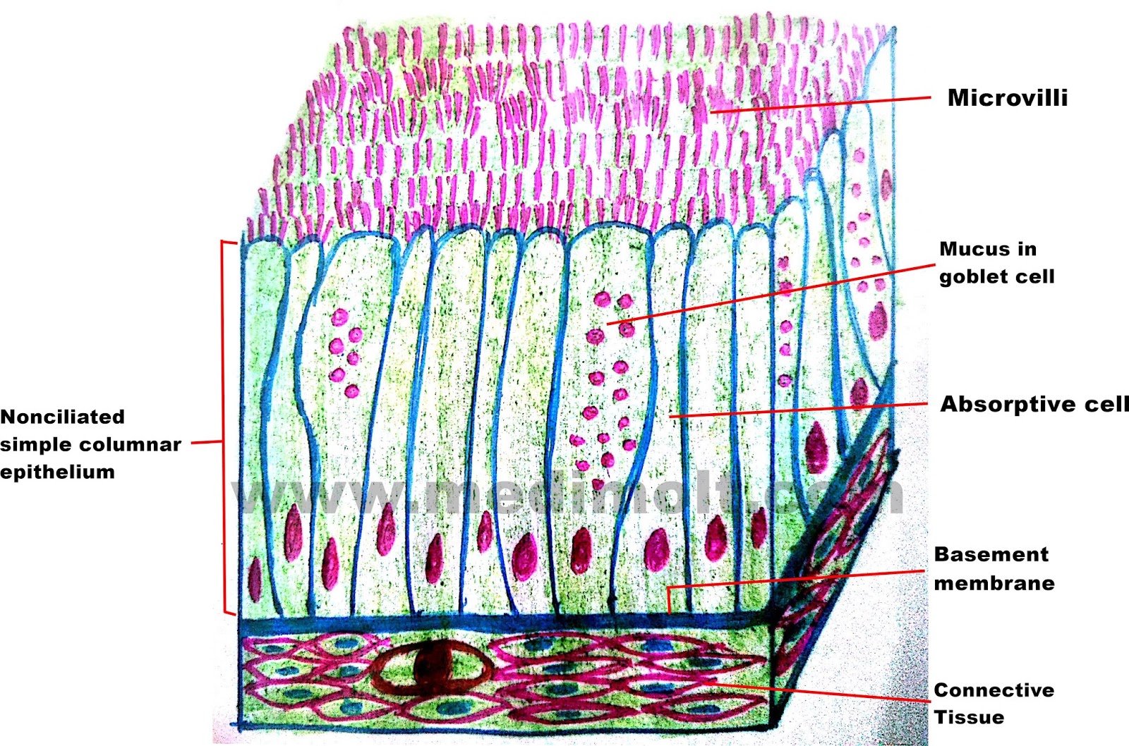
Histological structure pseudostratified columnar epithelium pseudostratified columnar epithelium is a single layer of ciliated, irregularly shaped cells containing many goblet cells.
Muscle tissue lumen basement membrane connective tissue epithelial tissue. Anatomy and physiology questions and answers. Seminal vesicle (ox) 1 2. Stratified squamous epithelia consist of multiple layers of cells with the outer most layer being squamous.
(3) squamous, thin, flat cells that look like the scales of a fish from a superior view.
It is mostly responsible for protection and mucous secretion. Start studying anatomy tissues labeling. Not involved in significant absorptive or secretory activity present in: Definition of stratified cuboidal epithelium.
Identify the structure labeled 2.
This type of epithelia lines the small intestine where it absorbs nutrients from the lumen of the intestine. The stratified cuboidal epithelium is composed of multiple layers of cells, in which the topmost layer is composed of cuboidal cells, while the lower layer could be columnar or cuboidal. March 23, 2022 stratified columnar epithelium may be found in the pharynx. Ciliated epithelium is a thin tissue that.
Simple columnar epithelia are also located in the stomach where it secretes acid, digestive enzymes and mucous.
Identify the structure labeled 3. This diagram represents pseudostratified columnar epithelium. Simple columnar epithelium features label the features of simple columnar epithelium. 5 the early primitive endodermal stratified columnar epithelium of the esophagus goes through multiple phenotypic expressions before it becomes lined entirely by stratified squamous epithelium.
As these ducts increase in diameter, their epithelium frequently becomes stratified columnar and eventually transitions to the stratified squamous epithelium of the vestibule into which they will empty.
There are three general shapes of epithelial cells: The modification of the cells on the apical surface is based on the location and function of. Pseudostratified columnar epithelium under a microscope this is not a true stratified epithelium but appears to be stratified. The cells of the epithelium are tightly packed, providing protection to surfaces of the.
Nucleus of a squamous cell outline the epithelium locations:
Columnar epithelium with goblet cells tissue / organ: In usual slides the boundaries between epithelial cells are often not clearly seen but because of the shape and spacing of the nuclei, the epithelium can be identified. Two or more layers of cells indicate the tissue has strata (layers), and are called stratified. Label the features of stratified columnar epithelium.
The cells that comprise the epithelial membranes are variously shaped and are named accordingly.
It is because different cellular heights and nuclei are also placed at a different levels. The columnar epithelial cells are shaped like a column, with the height being greater than the width. But, in the pseudostratified columnar epithelium, the nuclei appear to be arranged into two or more layers. Identify the epithelial tissue labeled 1.
Ducts of sweat glands large ducts of exocrine glands anorectal junction 30.
A labeled diagram and functions epithelium is a tissue that lines the internal surface of the body, as well as the internal organs. You know the nuclei of the columnar epithelium lie in a row toward the basal part of the cell. Cilia columnar cell cuboldal cell goblet cell connective tissue nucleus microvilli basement membrane உனை ) photo victor p eroschenko.