Normally lined by single layer of mucinous columnar epithelium. Epithelium forms continuous sheets of cells that line internal surfaces and cover the external surface of the body. It is located in the conjunctiva inside the eyelids and areas of tissue transition.
Pin on Histology Epithelial
Epithelia are classified based on three criteria:
Refer to the diagram at the end of this chapter for the tissue orientation and.
Is found at the distal portion of excretory ducts of the pancreas, salivary and sweat glands as well as in the conjunctiva of the eye. The epidermis of the skin is stratified squamous epithelium. However, the epithelium is still classified as squamous based on the cells of the surface layer. It's difficult to see the basal lamina in the region of the dividing cells, in the basal layer.
You will find the smooth muscle fibers surrounding the ducts and sperms in the lumen of the duct.
The stratified columnar epithelium has multiple layers of cells in which the apical layer is made up of columnar cells while the deeper layer can be either cuboidal or columnar. The pseudostratified stratified columnar epithelium lines the ductus epididymis. This is an example of thin skin. Transitional epithelium location and characteristics
In slide 29 and slide 176, this type of epithelium lines the luminal (mucosal) surface of the small and large intestines, respectively.
Click again to see term 👆. Thick layers of skeletal muscle is present at the level of the external urethral sphincter. It is most often found in the ducts from exocrine glands. In stratified squamous epithelium, the cells at the basal layer are cuboidal or even columnar.
It is not present in the larynx or pharynx.
You may identify stratified columnar epithelium histology slide with the following identifying characteristics under light microscope. Histology hint from sarah bellham: Has two or more layers of cells. Stratified columnar epithelium is often found at the junction between simple columnar epithelium and stratified squamous epithelium.
In some tissues, a layer of.
The modification of the cells on the apical surface is based on the location and function of. The ducts of the salivary glands fit into this category and are shown in this slide. An epithelium is the type of tissue that covers surfaces, usually the linings of hollow organs in the body, or in the case of the skin, the outer surface of the body. Presence of several layers of cells that rest on the basement membrane.
These ducts range from simple cuboidal, simple columnar, to stratified cuboidal epithelia.
The cornea is covered by a non. Such transitions occur in the ducts of larger glands. What appears stratified is, however, a single layer. The cells of the basal layer are cuboidal, while the cells at the surface are columnar, thus the name stratified columnar.
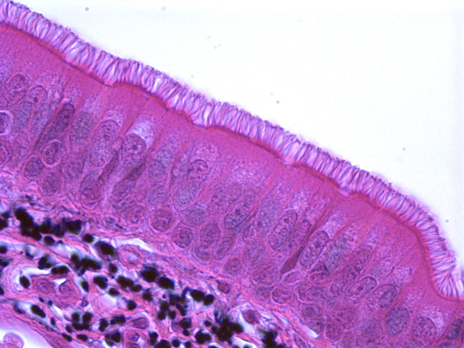
This section of pseudostratified columnar epithelium is from the mucosal lining of the trachea has a ciliated surface.
In the vas deference histology slide, you will find a. The epithelium classifies as pseudostratified; This type of epithelium is protective against chemical and mechanical damage, and water loss, and is found in skin, and oral epithelia. Stratified columnar epithelium is relatively rare, occurring mainly where simple epithelium transitions to a thicker stratified epithelium;
Respiratory epithelium is ciliated pseudostratified columnar epithelium found lining most of the respiratory tract;
It most often has only two layers of. Click here for answers and detailed explanations. Though it is a single layer of cells along the basement membrane, the alignment of the nuclei is not in the same plane and appears as multiple layers. The lining of the esophagus is stratified squamous epithelium.
The nuclei nearer the surface belong to the taller ciliated cells of the epithelium, while the deeper situated.
There are three principal shapes of epithelial cell: Is stronger than simple epithelium. In many cases, adjacent epithelial cells are linked by tight junctions so that the epithelium forms a barrier that regulates the movement or substances. For each histology question, pick the one best answer.
These can be arranged in a single layer of cells as simple epithelium, either squamous, columnar, or cuboidal, or in layers of two or more cells deep as stratified (layered), or compound, either squamous, columnar or cuboidal.
Notice the classic layering of the nuclei within the epithelium that give it the stratified appearance. Anatomic opening of the endocervical canal onto the ectocervix. Excretory duct, submandibular gland, 150x. Slide 29 (small intestine) view virtual slide slide 176 40x (colon, h&e) view virtual slide remember that epithelia line or cover surfaces.
As in the case of other stratified epithelium, the cells in the deeper layers might be different than the layer on the top.
Anatomic widening of the endocervical canal as it opens and gradually transitions into the uterine cavity. A basement membrane separates an epithelium from the underlying connective tissue. As these ducts increase in diameter, their epithelium frequently becomes stratified columnar and eventually transitions to the stratified squamous epithelium of the vestibule into which they will empty. It is a selective barrier that protects tissues and is often involved in absorption or secretion.
The urothelium changes into pseudostratified columnar epithelium.
This histology test bank is also useful for the histology questions on the usmle (usmle step 1).