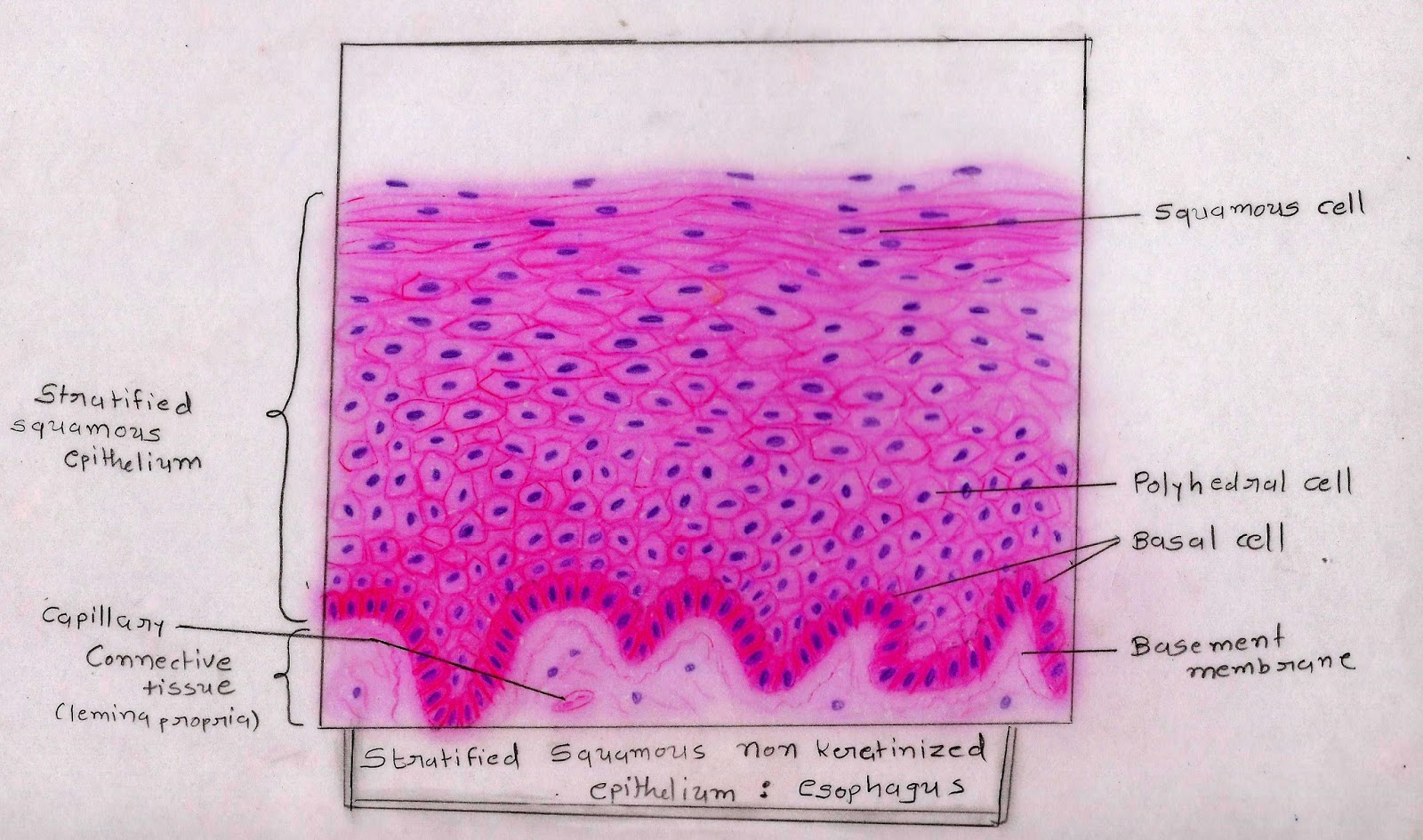
Find this pin and more on file diagrams by histopedia. (a) stratified squamous epithelium of the first esophageal region in. Identify the type of epithelium indicated by the.
Stratified epithelia Atlante di Istologia Histology Atlas
The cells in the apical layer and several layers deep to it are squamous while the cells in deeper layers vary from cuboidal to columnar.
Separated from underlying connective tissue by a basement membrane.
Apical surface is the free surface. An overview of epithelium, including example of six different kinds of epithelium: Little intercellular material is present so cells are closely apposed. Stratified squamous epithelium under a microscope
What type of tissue is this?
Epithelial tissue histology slide with microscopic images and labeled diagrams. However it has been shown that all of the cells of a pseudostratified epithelium are in contact with the basement membrane bm. These labelled diagrams should closely follow the current science courses in histology, anatomy and. It is because different cellular heights and nuclei are also placed at a different levels.
Pseudostratified epithelium is also sometimes referred to as respiratory epithelium, since ciliated pseudostratified columnar epithelia is mainly found in the larger respiratory airways of the nasal cavity, trachea and bronchi.
These labelled diagrams should closely follow the current science courses in histology, anatomy and. Stratified squamous moist epithelium lining the esophagus changes abruptly to a simple columnar epithelium forming the sheet gland of the stomach. What appears stratified is, however, a single layer. This section of pseudostratified columnar epithelium is from the mucosal lining of the trachea has a ciliated surface.
Notice the classic layering of the nuclei within the epithelium that give it the stratified appearance.
The pseudostratified columnar epithelium comprises a single layer of cells but seems to be multilayered. Simple squamous, simple cuboidal, simple columnar, stratified squamous, tr. Slides 27*, 125*, 110, and 73* the larger conducting passageways of the respiratory system, the trachea and the bronchi, are lined by pseudostratified ciliated columnar epithelium. Contain junctional complexes allowing for adhesion and communication.
In a pseudostratified epithelium one can see nuclei n at several different levels, giving an appearance of an epithelium composed of layers of cells (a stratified epithelium).
The term pseudo (false) reminds one that this is not a stratified epithelium. The pseudostratified stratified columnar epithelium lines the ductus epididymis. To locate bronchi on slide. On the other diagram, you will see the transverse section of the simple columnar epithelium.
Columnar epithelium with goblet cells tissue / organ:
You may know the details of histological features of the simple columnar epithelium from another article by anatomy learner. Columnar epithelium with goblet cells tissue / organ: You will find the smooth muscle fibers surrounding the ducts and sperms in the lumen of the duct. Move substances through the lumen.
Simple columnar epithelium (diagram) stratified columnar epithelium (diagram) ciliated pseudostratified columnar epithelium (diagram) transitional epithelium (diagram) simple squamous epithelial tissue (image).
The nuclei nearer the surface belong to the taller ciliated cells of the epithelium, while the deeper situated nuclei. Again, in the lingual and tubal tonsil, the lining epithelium is the nonkeratinized stratified squamous epithelium. Composed of sheets or layers, or clusters of closely alined cells. In the vas deference histology slide, you will find a.
If you want to memorize them, you may read this article from anatomy learner.
I hope you have a good piece of knowledge on the histology of nonkeratinized stratified squamous epithelium and pseudostratified columnar epithelium. Pseudostratified columnar epithelium under a microscope with a labeled diagram.
:background_color(FFFFFF):format(jpeg)/images/article/en/stratified-epithelium/kxsJZMc6RS4j61ethfEOw_A4WBRjTb2LJJRtmn2wVxCw_stratified_squamous_epithelia02.png)

:background_color(FFFFFF):format(jpeg)/images/library/2348/e4C4QQTQQqT43OdGZOidHQ_simple_cuboidal_epithelium_01.png)


