Simple epithelium has only one layer of cells, while stratified epithelium has two or more layers. About press copyright contact us creators advertise developers terms privacy policy & safety how youtube works test new features press copyright contact us creators. Huge collection, amazing choice, 100+ million high quality, affordable rf and rm images.
Free Free Commercial Use Clipart, Download Free Clip Art
In humans, a simple columnar epithelium lines most organs of the digestive tract including the stomach,.
The stratum basale (also known as stratum germinativum) is the inner layer of the epidermis.
Stratified columnar epithelium can be found in the urethra. Based on structure, and based on form and function. Keratinizing stratified squamous epithelium tissue locations function & notes drawing specific location: Epithelium is a tissue that lines the internal surface of the body, as well as the internal organs.
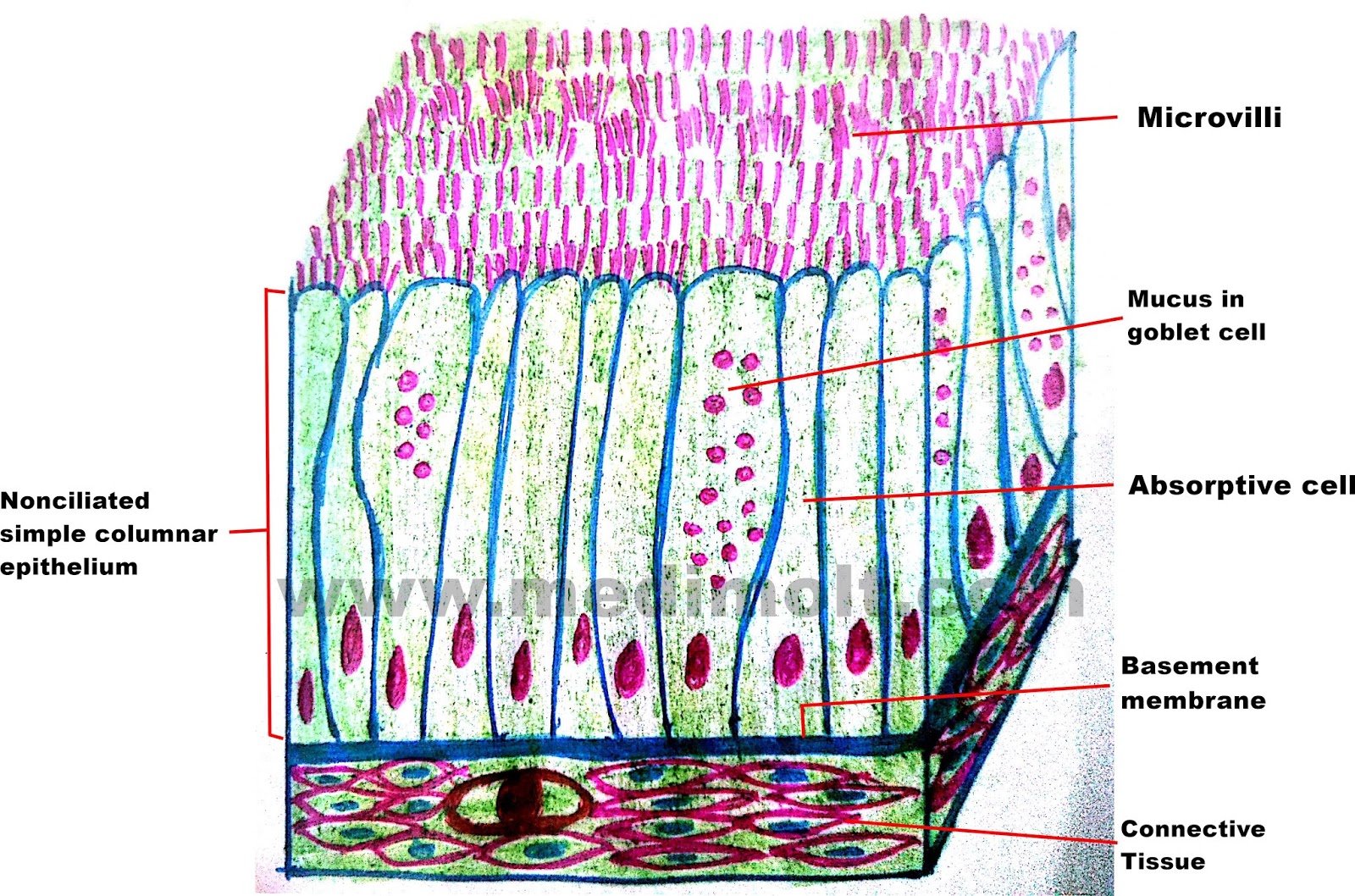
On a blank piece of paper draw the characteristics of columnar epithelium.
Draw diagram of each type of epithelial tissue. As in the case of other stratified epithelium, the cells in the deeper layers might be different than the layer on the top. Od cent stratified epithelium, stratified squamous epithelium draw an example. Simple epithelium is one of the types of epithelium that is divided into simple columnar epithelium, simple squamous epithelium, and simple cuboidal epithelium.
In our drawing and in the histological sample, indicate the goblet cells, which secrete mucus to trap.
First, draw a section of simple columnar epithelium, which comprises a basement membrane supporting cells that are taller than they are wide; Upload your drawing to the annotate panel. It is mostly responsible for protection and mucous secretion. Bodytomy provides a labeled diagram to help you understand the structure and function of simple columnar.
It is located in the conjunctiva inside the eyelids and areas of tissue transition.
Enter the important histological characteristics of columnar epithelium into the table. You know the nuclei of the columnar epithelium lie in a row toward the basal part of the cell. It is because different cellular heights and nuclei are also placed at a different levels. Columnar epithelium with goblet cells tissue / organ:
Notice how the simple columnar cells lining the uterine cervix transitions to stratified squamous epithelium.
Stratified tissues are named based on the shape of the most apical cells. The pseudostratified columnar epithelium comprises a single layer of cells but seems to be multilayered. The principle structural types are simple and stratified. Pseudostratified columnar epithelium under a microscope this is not a true stratified epithelium but appears to be stratified.
Epithelial tissue is classified in two ways:
But that of pseudostratified columnar epithelium appear to be arranged in two or more layers giving the impression that the epithelium is more than one cell thick but it is not the reason for this is that some cells are broader near the base while others are broader near the apex and. Use colored pencil and label main structures 4. Pseudostratified columnar epithelium under a microscope with a labeled diagram. Location function description simple epithelium, simple columnar ciliated draw an example.
Brush border & terminal bars.
With oval and basal nuclei. Nonkeratinizing stratified squamous epithelium tissue locations function & notes drawing 2. Click on the button or title to get to the appropriate image: Make sure you include the details you entered into the table in your drawing.
The stratified columnar epithelium has multiple layers of cells in which the apical layer is made up of columnar cells while the deeper layer can be either cuboidal or columnar.
Which of the following tissue description a) flat cells with irregular boundaries. The modification of the cells on the apical surface is based on the location and function of. The name of the specific tissue is simple columnar epithelium. A typical example of stratified squamous keratinized epithelium is the epidermis.
So an epithelium that has several layers and the apical cells are flattened and scale shaped, the name of the specific tissue is stratified squamous epithelium.
But, in the pseudostratified columnar epithelium, the nuclei appear to be arranged into two or more layers. The drawings of histology images were originally. In the usual columnar epithelium the nuclei lie in a row, towards the bases of the cells; Learn vocabulary, terms, and more with flashcards, games, and other study tools.