It produces melatonin, a serotoninderived hormone, which affects the modulation. Development of the pineal gland: It is attached by a stalk to the posterior wall of third ventricle.
Brain, coronal, labelled Radiology Exam Pinterest
It was once known as “the third eye”.
The pineal gland is a midline structure, located between the two cerebral hemispheres.
Also, mri can aid in the detection of tumor seeding to other parts of the nervous system. What are the grades of pineal region. The gland is a conical, grey body measuring 5 to 8mm in length and 3 to 5mm in its greatest width. As such an understanding of the normal anatomy of the region as well as a systematic approach to.
Pineal gland also called epiphysis cerebri.
The visualization of cp is difficult due to its location and surrounding structures and so far, no standardized method exists. The pineal gland (cp) is located centrally in the brain and produces melatonin. The pineal gland is a neuroendocrine organ that comprises a part of the epithalamus, one of the three divisions of the diencephalon.other components of the epithalamus are the stria medullaris, habenular nuclei, posterior commissure and paraventricular nuclei. Cysts and concrements are frequent findings on mri but their significance is still unclear.
These tumors begin in the brain (in the pineal gland) but can spread to the spinal cord.
Overview • in human the cycles of sleep and wakefulness usually mimics the rhythm of light and dark. The normal size ranges between 10 and 14 mm [].it is commonly located along the midline above the superior colliculi and inferior to the splenium of the corpus callosum. Place the cursor over the image to display the anatomy revised 06/25/04. The pineal gland, (pineal body ;
Reduced resolution compared to mri.
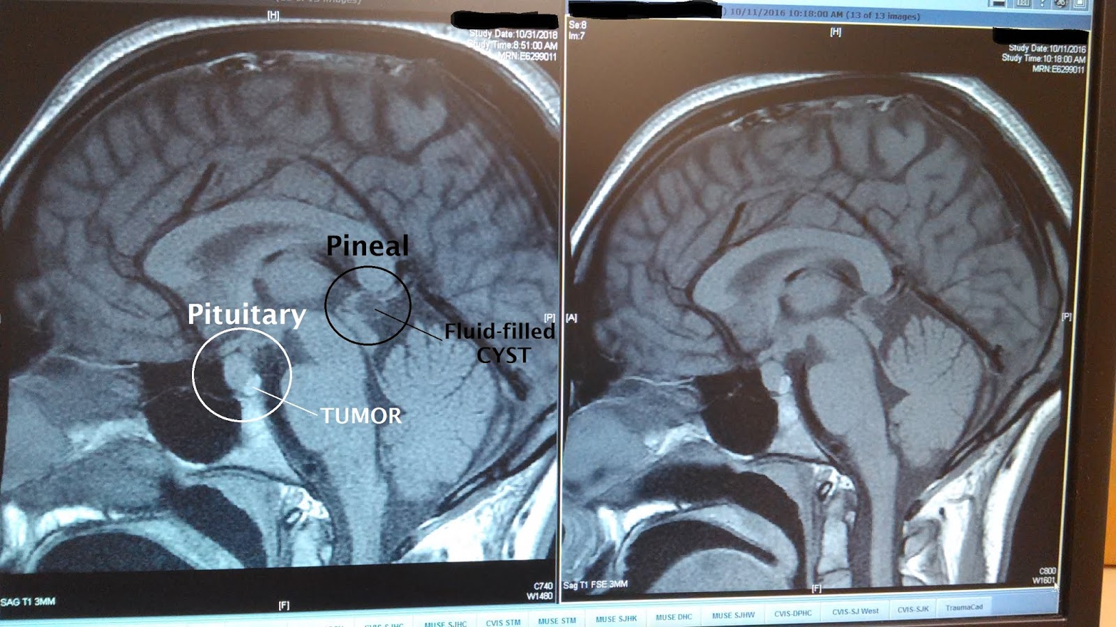
Pineal region tumors are primary central nervous system (cns) tumors. Drawing showing the anatomy of the pineal gland and pituitary gland in the brain. This gland is rich in calcium levels. To get an accurate diagnosis, a piece of tumor tissue will be removed during surgery, if possible.a neuropathologist should then review the tumor tissue.
The electronic curriculum is copyrighted 1998,.
(1) pineal gland, (2) splenium of the corpus callosum, (3) third ventricle, (4) tegmentum of the midbrain, and (5) tectum of the midbrain. In length which lies in the depression between the superior colliculi. An mri study made in iceland shows that the median volume of the pineal gland is 207 mm3 and 59% of the glands have presented cysts and 20% have presented calcifications. Harnsberger hr, osborn ag, ross js, moore kr, salzman kl, carrasco cr, halmiton be, davidson hc, wiggins rh.
Magnetic resonance imaging (mri) with enhancement provides exquisite anatomic detail, outlining the lesion, the cerebrospinal fluid (csf) pathways, and the venous anatomy around the vein of galen.
It develops as an outgrowth from the third ventricle of the brain. The pineal gland produces the hormone melatonin, and its volume may influence melatonin levels. The pineal gland is a pine cone shaped gland of the endocrine system. Brain, head and neck, spine.
Autopsy studies have shown that the average size of the pineal gland is similar to that of a grain of rice.
A systematic approach to the pineal region is crucial as it is at the confluence of many intracranial structures/regions and is the site of origin of a number of unique pathologies as well as playing host to many entities which are more frequently encountered elsewhere. This article will explore the anatomy. It is the major site for melatonin secretion, which regulates the body’s internal clock (circadian rhythm). However, as with ct, the mri characteristics of pineal region tumors are usually.
Brain, anato my created date: