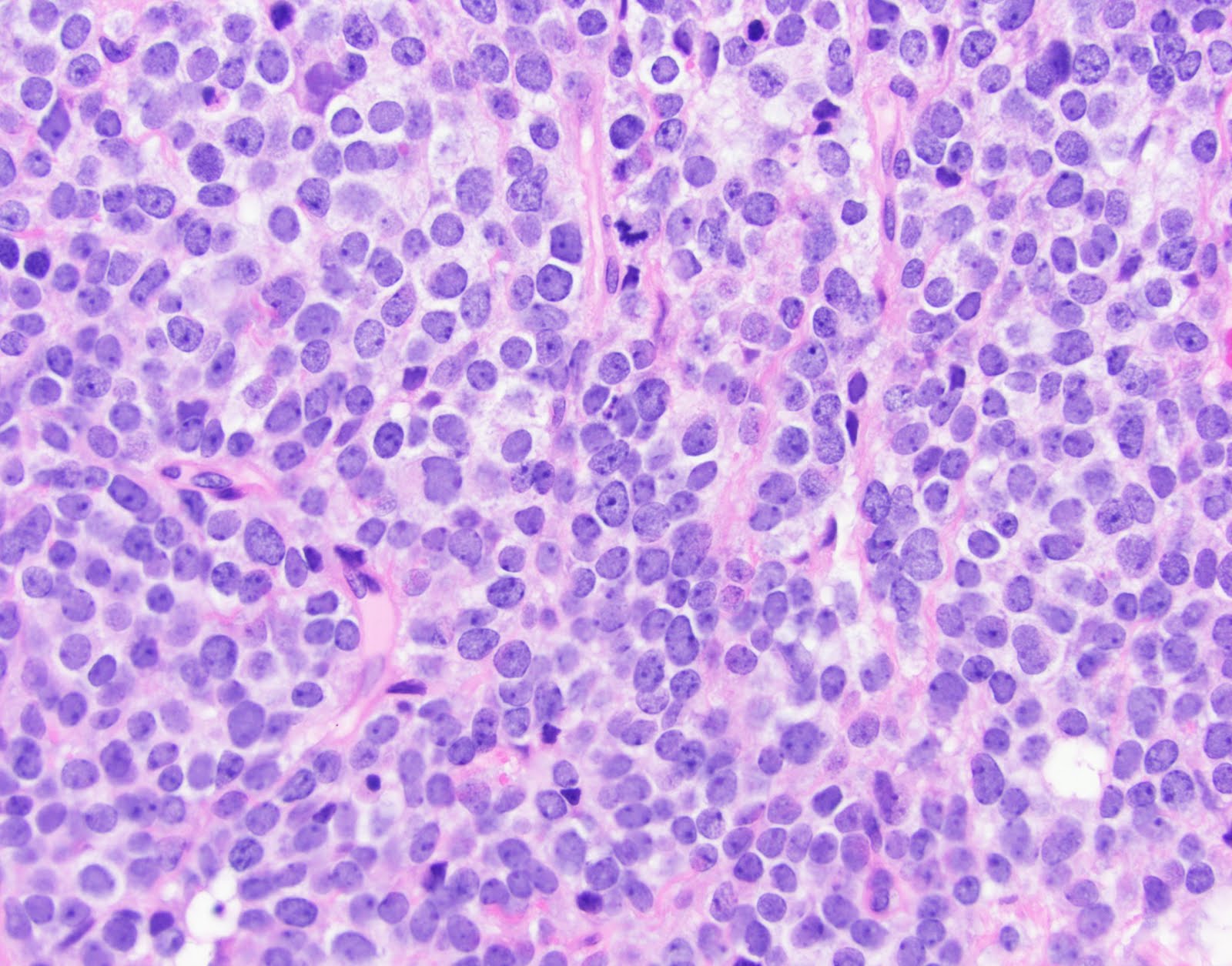
The pineal gland or epiphysis cerebri is a small neurosecretory organ in the brain containing secretory neurons (pinealocytes), interstitial glial cells, and axons that communicate with other parts of the brain. The pineal gland regulates daily body rhythms. Brain sand surrounded by pinealocytes low magnification high magnification.
"Pineal Gland Histology" Drawstring Bag by deltoid Redbubble
Germinoma ~ 50% of (pineal) tumours.
Pineal gland also called epiphysis cerebri.
The gland is a conical, grey body measuring 5 to 8mm in length and 3 to 5mm in its greatest width. Posterior lobe of pituitary gland. Pineal parenchymal tumor of intermediate differentiation. This article will explore the anatomy.
These features correlate closely with what is seen on histology.
A high magnification shows connective tissue septa dividing the pineal gland into lobules. Introduction to histology of the pineal gland, as presented by the university of rochester pathology it program. Pineal gland lacks a blood brain barrier; This gland is rich in calcium levels.
Pineal gland anatomy of pineal gland histology of pineal gland 1) pinealocytes 2) astrocytes physiological function references 3.
Histology slide courtesy of mt. The pineal gland has a predilection for calcification which is invariably histologically present in adults but rarely seen below the age of 10 years 6. Teratoma ~ 15% of tumours. Normal histology see neurohistology#pineal gland.
In some of these glands only small islands of parenchymal cells surrounded by connective tissue.
Cone shaped structure attached by a stalk to roof of the diencephalon; The pineal gland consists of several types of cells, principally pinealocytes and astrocytes. Cytologic features of the pineal gland are peculiar when compared to other normal structures of the central nervous system. The pineal gland is made up of follicules and lines of pinealocytes and glial tissue.
The pineal gland the function of this small organ near the center of the mammalian brain has long been a mystery.
Recent studies indicate that it is a biological clock that regulates the activity of the sex glands by richard j. In an adequate clinical context, and in combination with frozen sections, cytology allows a specific recognition of the pineal gland during intraoperative. It is the major site for melatonin secretion, which regulates the body’s internal clock (circadian rhythm). Variations in weight are present only.
Develops at month 2 of gestation as diverticulum in diencephalic roof of third ventricle.
It is an important endocrine gland in. Thyroid gland and parathyroid gland. The remaining cell population consists of interstitial cells, which are modified astrocytes similar to pituicytes in the neurohypophysis. Between superior colliculi at base of brain;
The pineal gland is a pine cone shaped gland of the endocrine system.
Some glands have a well defined, lobular structure, strands of connective tissue separating the groups of parenchymal cells. It was once known as “the third eye”. Also called epiphysis, pineal body. It sits in the groove between the two superior colliculi, and is bilaterally.
The overall architecture of the pineal gland is extremely variable.
Replaced by connective tissue after puberty. The capsule of the gland is well. Pineal recess communicates with the 3rd ventricle in the adult; In others the connective tissue is more extensive and the bands are wider.
Develops from the roof of the diencephalon (the epiphysis);
Connective tissue septa penetrate the gland, subdividing it into indistinct lobules. It follows the circadian rhythms set by the suprachiasmatic nucleus of the hypothalamus. Pituitary gland anterior lobe posterior lobe. Histologically, pinealocytes have a slightly basophilic cytoplasm with large irregular or lobate.
Anterior lobe of pituitary gland.
The histology of the pineal gland in odocoileus virginianus terry michael de villiers eastern illinois university this research is a product of the graduate program inzoologyat eastern illinois university.find out more about the program. The gland is surrounded by a capsule of pia mater and arachnoid elements; It is photosensitive and receives its signals from the retinohypothalamic tract. The pineal gland, also called the pineal body, develops as an outward projection from the posterior wall of the third ventricle, below the splenium of corpus callosum.
Describe the development of the pineal gland.
Wurtman and julius axelrod buried nearly in the center of the brain in any mammal is a small Some glands have a perfectly lobular aspect, separated by connective tissue, in others the connective tissue is much more abundant, and the parenchyma is arranged in a insular pattern.


