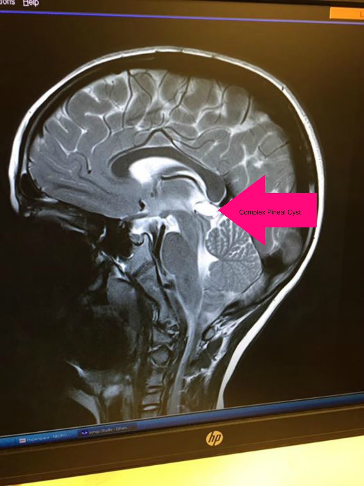
A pineal cyst is a growth with smooth sides that appears on or near the pineal gland. As a general rule, pineal cysts are described as such when they reach a size of at least 5 mm, but there are reports of pineal cysts less than 2 mm in diameter. Symptoms of a pineal cyst.
Brain Pathology Pineal Cyst Mri Exam Stock Illustration
It’s caused when the cyst blocks the flow of csf in the brain.
The pineal gland increases in weight and volume with age and can even gradually increase in adulthood.
Hydrocephalus can cause symptoms such. An mri of my pituitary gland last week revealed a 13mm simple nonenhancing pineal cyst. A total of 178 patients (63.3%) were female, and the age at diagnosis ranged from 16 to 84 years. Mri findings the cyst contents are hypointense on t1w images and hyperintense on t2w images.
A remnant of the pineal diverticulum or distension of its obliterated portion has been postulated as a possible source of pineal cyst.
Pineal cysts (pc) are cysts which are frequently detected incidentally in brain magnetic resonance imaging (mri). They may be isointense or hyperintense on pdw and flair images, depending on the protein content of the. I began having problems 7 years ago and had a couple mri's each showing a pineal gland cyst, and optic neuritis. In rare cases, a pineal cyst may cause csf to build up on the brain.
In most cases, a pineal cyst is a benign tumor which does not cause any symptoms.
These cysts are benign, which means not malignant or cancerous. In some patients it will be recommended to follow the cyst with mri to determine if it is growing. The classic version of the pineal cyst is usually small (up to 10 mm) and one chamber. I have suffered over the years but recently had a terrible dizzy spell lasting 2 days.
It is a benign cyst involving the pineal gland usually measuring less than 1 cm with no enhancing nodular, solid component.
Histologically, they are composed of an inner layer of gliotic tissue, middle layer of pineal parenchymal tissue and an outer layer of connective tissue. This has mildly greater signal than csfon t1 and flair and similar to csf on t2. I've posted about this before but it's been a little while. The principal indication for head mri was headache (50.2%), although no symptoms were deemed attributable to pineal disease.
The pineal gland develops by the proliferation of walls of the third ventricle diverticulum in the diencephalic roof.
In series of magnetic resonance imaging (mri) studies, the prevalence of pineal cysts ranged between 1.3% and 4.3% of patients examined for various neurologic reasons and up to 10.8% of asymptomatic healthy volunteers. This case illustrates the appearances of a large pineal cyst with fluid which does not fully attenuate on flair. Occasionally a cyst may enlarge and press on surrounding structures causing symptoms such as headaches and blurred vision. Pineal cyst discovered in mri for pituitary gland.
Such a growth can cause symptoms ranging in severity from nausea and headaches to coma.
Incidentally detected on mri for sellar tumour. A total of 281 patients were identified with pineal cysts. At times, the pineal gland can appear prominent and simulate an enhancing lesion, with or without the presence of a cyst. This sort of lesion needs followup.
This is usually because the cyst has grown so much that it has pressed into other parts of the brain.
Pineal cysts (pcs) are a benign lesion of the pineal gland that have been known to the medical community for a long time. Cells types adjacent to the pineal gland. My pituitary was being imaged for thyroid problems i've been experiencing for the past three months (fluctuating tsh leaning more toward hypo yet elevated rai uptake, a litany of symptoms like my neck being swollen and tight. Large cysts rarely cause symptoms of paralysis of upward gaze and headache because of mass effect on the tectum and hydrocephalus due to compression of the cerebral aqueduct.
In rare situations, the pineal cyst can be symptomatic.
It is not completly round in its configuration though appears to have a thin border. Pineal cysts occur in all ages, predominantly in adults in the fourth decade of life. The founder of modern pathology, rudolf virchow, described this entity as hydrops cysticus glandulae pinealis in 1865 [ 1 ], and campbell provided the first detailed description of its histological structure in 1899 [ 2 ]. Homogeneous cystic abnormalities of the pineal gland, called pineal cysts, are common incidental findings on brain mr imaging [1, 2].
Most pineal gland cysts have a characteristic appearance on an mri and can sometimes be distinguished from other tumors of the pineal region, such as pineoblastoma, pineocytoma and pineal germinoma.
If this occurs, the cyst can be removed using surgery. The median size of pineal cyst at diagnosis was 10 mm. A pineal cyst does not usually cause any symptoms. I saw the neurosurgeon on monday for the pineal cyst that was found on my mri last month.
The diagnosis of pineal cyst is usually established by mri.
In rare cases, extra csf or bleeding into the cyst may cause headache or make it hard to look up. Neurosurgeon says he doesn't think it's causing any of my issues and just keep treating for my lyme and recheck it in 6 months.


