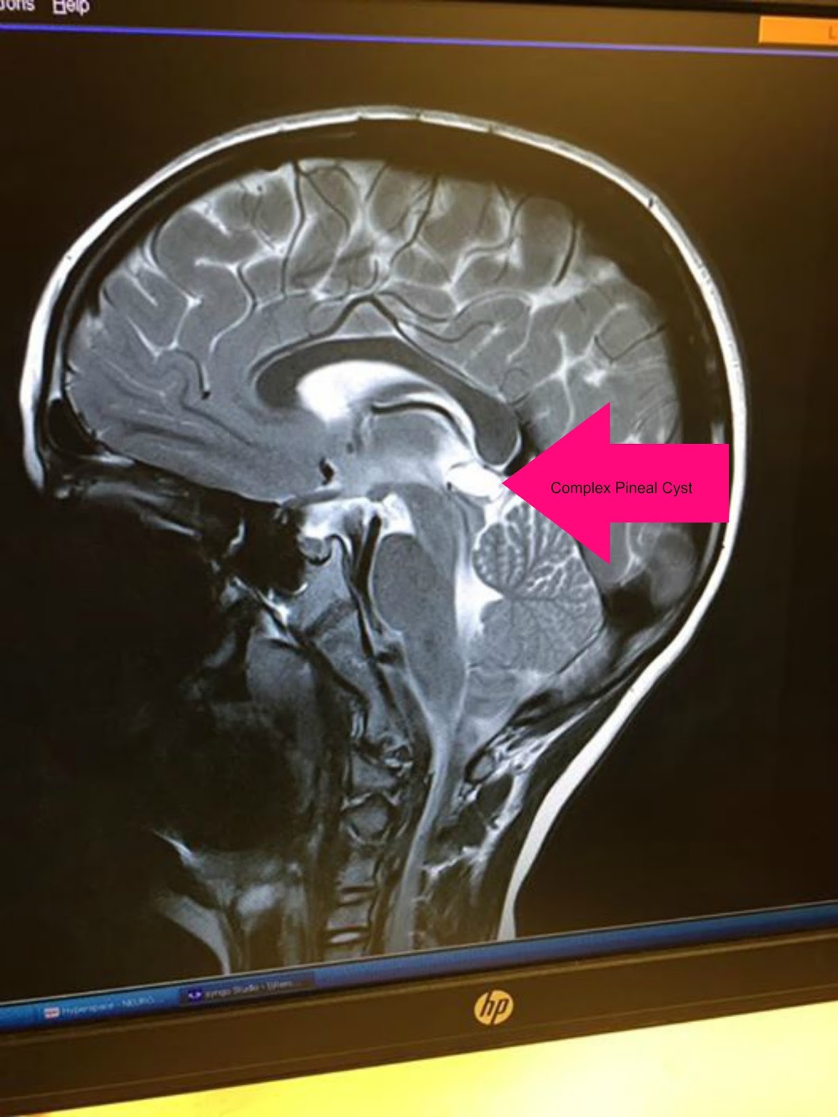
True pineal gland cysts are not brain tumors; Pineal cysts are small lesions usually asymptomatic and encountered incidentally. In series of magnetic resonance imaging (mri) studies, the prevalence of pineal cysts ranged between 1.3% and 4.3% of patients examined for various neurologic reasons and up to 10.8% of asymptomatic healthy volunteers.
Pineal cyst Radiology Case
The pineal cyst is a rare cystic mass.
It is characterized by premature aging.
This disease rarely has serious consequences if the cyst of the pineal gland does not begin to grow sharply. These cysts are benign, which means not malignant or cancerous. The diagnosis of pineal cyst is usually established by mri with defined. A pineal cyst is usually found by chance on an imaging test of the brain.
The large majority of pineal gland cysts do not enlarge much if at all.
Mri scans of your brain may be done to get more. Pineal cysts should be considered for surgery only when the diameter of the cyst is more than 1.0cm, or when obstructive hydrocephalus is verified on mri or ct, and/or when definitive neurological signs such as parinaud's syndrome are present. Found a frequency of 23% in brain scans. Clinical presentation in the emergency setting:
Ct of pineal region tumors the computed tomographic (ct) features of pineal region tumors were analyzed in 60 histologically proven tumors.
This is the largest reported series of histologically verified pineal region tumors. This is the largest reported series of histologically verified pineal region tumors studied with ct. Peripheral contrast enhancement is characteristic of the predominant number of cysts, and a band of calcifications (border) is observed in approximately every. This may be one of the following:
A very rare genetic disorder caused by mutations in the lmna gene.
Most pineal cysts are found incidentally during mr screening studies and no surgical interventions are needed. It often has concentric calcification involving the wall of the cyst. Rather, they are a completely benign cyst. 3) radiologists cannot easily distinguish between cysts and benign tumors, often leading to misdiagnosis.
This sort of lesion needs followup.
Found a frequency of 23% in brain scans (with a mean. What kind of pineal region tumors are there? However, some enlarge over time slowly. A pineal gland cyst is a usually benign cyst in the pineal gland, a small endocrine gland in the brain.
Signs and symptoms include failure to thrive, limited growth, alopecia, wrinkled skin, small face, development of atherosclerosis, and heart disease.
This may be one of the following: A disease that produces rapid aging, beginning in childhood. [1] a 2007 study by pu et al. A benign tumor is not metastatic, not malignant.
The pineal gland increases in weight and volume with age and can even gradually increase in adulthood.
A pineal cyst is usually found by chance on an imaging test of the brain. This test uses large magnets and a computer to create images of the body. Pineal cysts occur in all ages, predominantly in adults in the fourth decade of life. 2) pineal cysts can be symptomatic if they are larger than 0.5 cm.
The cause of these cystic masses are not known.
Problems occur when the cysts cause compression in the brain, or when they are associated with apoplexy or hydrocephalus. This case illustrates the appearances of a large pineal cyst with fluid which does not fully attenuate on flair. The vast majority of pineal cysts are small (ct</strong> scans or mri. A 2007 study by pu et al.
According to statistics, only 1.5% of patients with signs of a brain disease are diagnosed with this type of cyst.
A cyst is a benign formation that can change its size. It is a benign cyst involving the pineal gland usually measuring less than 1 cm with no enhancing nodular, solid component. Sometimes an mri of the pineal cyst needs to be repeated with an. Despite the pineal gland being in.
Larger cysts can present with mass effect on the tectal plate leading to compression of the superior colliculi and parinaud syndrome.


