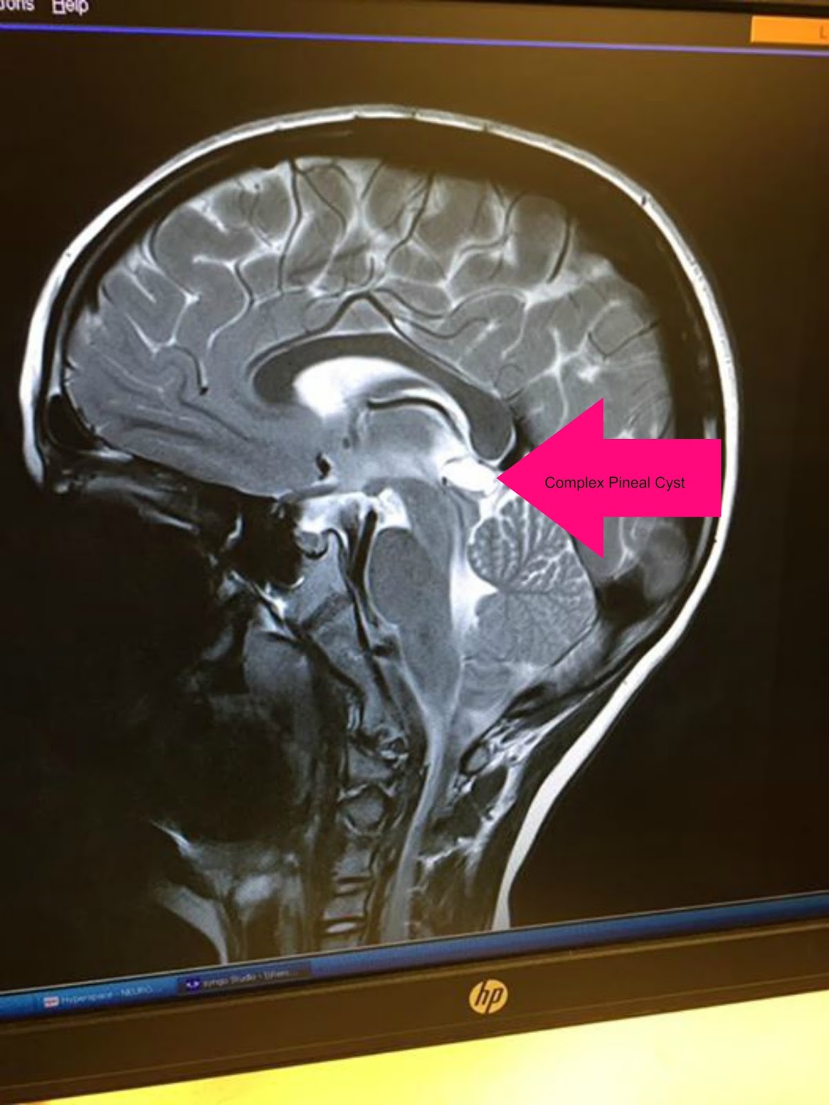
The pineal gland is part of the epithalamus. Anderson published ct studies in 2006 that have shown that the incidence of calcifications in the pineal gland has risen, one study presents rare calcifications in children, the youngest being 3 years old. These tumors begin in the brain (in the pineal gland) but can spread to the spinal cord.
11 Simple Pineal Cyst Radiology Key
Develops at month 2 of gestation as diverticulum in diencephalic roof of third ventricle.
Now that you have learned the anatomy, do the labeling activity below.
Anterior horn of the lateral ventricle A structure of the diencephalon of the brain, the pineal gland produces the hormone melatonin. It is very commonly partly or fully calcified in adults Computed tomography (ct) scans can better show calcification than mr imaging (mri).
It was once known as “the third eye”.
Anthony james doyle and graeme d. Pineal region tumors are primary central nervous system (cns) tumors. Genu of the corpus callosum. Replaced by connective tissue after puberty.
Harnsberger hr, osborn ag, ross js, moore kr, salzman kl, carrasco cr, halmiton be, davidson hc, wiggins rh.
This is the largest reported series of histologically verified pineal region tumors studied with ct. Recently, radiological studies of the pineal gland have been mainly conducted by ct on the existence of pineal calcification and the proportion of calcification on axial ct images over different populations [3,4,5,6, 27]. Diagnostic and surgical imaging anatomy: Head ct > anatomy > normal anatomy 6.
Click image to align with top of page.
It is attached by a stalk to the posterior wall of third ventricle. On the image below, locate the following anatomy: It is the major site for melatonin secretion, which regulates the body’s internal clock (circadian rhythm). Hover on/off image to show/hide findings.
The pineal gland further performs a function in the regulation of female hormone levels, and it may influence.
The midline supracerebellar route is the shortest and provides direct exposure of the pineal gland, although the culmen and inferior and superior vermian tributaries. The pineal gland is located immediately posterior to the third ventricle; Tap on/off image to show/hide findings. The pineal gland is a midline structure, located between the two cerebral hemispheres.
The pineal gland, also called the pineal body, develops as an outward projection from the posterior wall of the third ventricle, below the splenium of corpus callosum.
Between superior colliculi at base of brain; Brain, head and neck, spine. Twenty ct angiograms were examined to measure the depth of the pineal gland, slope of the tentorial surface of the cerebellum, and angle of approach to the pineal gland in each approach. It sits in the groove between the two superior colliculi, and is bilaterally.
What are the grades of pineal region.
Ct of pineal region tumors the computed tomographic (ct) features of pineal region tumors were analyzed in 60 histologically proven tumors. The pineal gland is composed of cells called pinealocytes and cells of the nervous system called glial cells. Also called epiphysis, pineal body. To get an accurate diagnosis, a piece of tumor tissue will be removed during surgery, if possible.a neuropathologist should then review the tumor tissue.
The pineal gland, also known as the epiphysis cerebri, epiphysis or third eye is a small endocrine gland in the brain.
Axial section 6 is approximately 4 cm above the oml.


