The two palatine bones ( l., palatum “palate”) form portions of the hard palate, lateral walls of the nasal cavity, and floors of the orbits. It is an oblong, vertical plate, and possesses two surfaces, nasal and maxillary, and four. In humans, this bone is found between the maxilla, or upper jawbone, and the sphenoid bone, located at the base of the skull.
The Palatine Bone Anatomy, Borders and Development Kenhub
The palatine bones, like many bones in the human body, are paired bones named.
Os palatinum) is a thin paired bone that participates in forming the nasal cavity and oral cavity, as well as the pterygopalatine fossa.
The largest region of each of the palatine bone is the horizontal plate. Anterior view of the skull featuring the bones of the viscerocranium: Palatine bones are the most dorsal bones of the viscerocranium, acting as the bridge between processus pterygoideus & corpus ossis sphenoidalis from one side and maxilla from the other. They also form part of the floor and sides of the nasal cavity and extend to the orbit of the eye forming a small section of the orbit.
Useful notes on the palatine bones of human skull perpendicular plate:.
The cranium and the face (figs 10.1, 10.2 and 10.3 ). The horizontal laminae or plate of the palatine bone. Each palatine bone of a cat comprises a horizontal plate and a curved irregular verticle plate. Video about cranial bones and facial subdivisions of human skull.
The oral cavity, nasal cavity and the orbits.it does so by articulating with five bones;
It is located between the maxilla and sphenoid bone and is a part of inferior skull surface. A palatine bone of dog skull. It projects upward and laterally as an expanded process from. 10.1 position of the bones of the skull (anterior aspect).
Use this facial bone mnemonic list to remember the anatomy, names, and structure of each of the facial bones of the skull.
Palatine bone (os palatinum) the palatine bone is a paired bone located between the maxillae and the pterygoid process of the sphenoid bone.it participates in building the three cavities within the skull; About press copyright contact us creators advertise developers terms privacy policy & safety how youtube works test new features press copyright contact us creators. The palatine bones ex situ & in situ. The palatine bone is one of a pair of irregularly shaped bones that contribute small areas to the lateral walls of the nasal cavity and the medial wall of each orbit.
Each irregular quadrilateral horizontal plate of the palatine forms the caudal and medial part of the roof of the cat’s mouth.
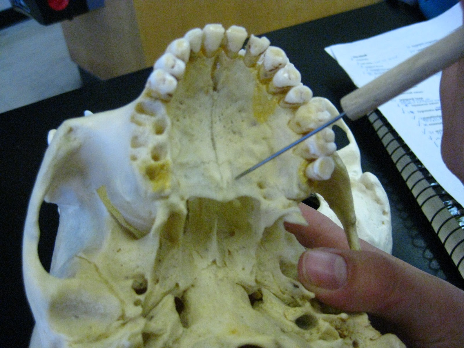
The skull can be divided into 2 sections: The plates from the right and left palatine bones join together at the midline to form the posterior quarter of the hard palate (see figure 6a). They also form part of the floor of the nasal cavity (the hard palate separates the oral cavity from the nasal cavity). The lateral margin of the flat palatine.
Uses labeled diagrams to show the structure and anatomy of each facial bone.
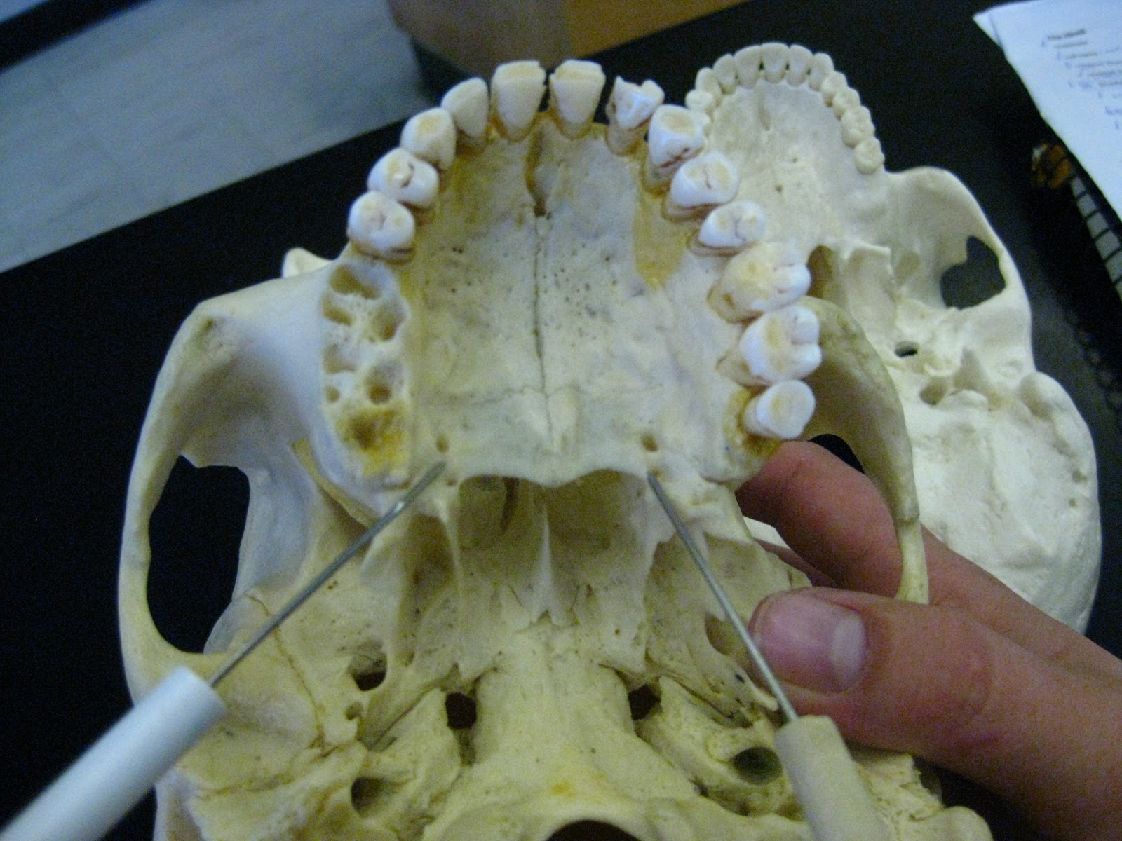
The greater palatine canal goes through the palatine and sphenoid bones, connecting the pterygopalatine fossa to the oral cavity.it transmits the descending palatine. The meaning of palatine bone is a bone of extremely irregular form on each side of the skull that is situated in the posterior part of the nasal cavity between the maxilla and the pterygoid process of the sphenoid bone and that consists of a horizontal plate which joins the bone of the opposite side and forms the back part of the hard palate and a vertical plate which is extended. Each bone primarily consists of a horizontal plate and a perpendicular (or. The palatine bone is one of the bones of the face.
Caudal to the palatine process of the maxilla, you will find the paired palatine bone in the cat skull anatomy.
The palatine bones are two small bones of the skull which together form the rearmost section of the hard palate (roof of the mouth within the oral cavity). They form the hard palate with the maxillary bones. This location leaves the palatine bone lying at the back of the nasal cavity. The greater palatine foramen is a small opening in the horizontal plate of the palatine bone which serves as a passage for the greater palatine nerve coming out from the greater palatine canal into the oral cavity.
The nasal cavity is a space filled with fluid and is found in the middle of the face, just behind the nose.
This palatine bone of the dog is divided into horizontal and perpendicular laminae. Maxilla, sphenoid, ethmoid, inferior nasal concha, and vomer. It is located between the maxilla and sphenoid bone and is a part of inferior skull surface. The palatine bone locates caudomedial to the maxilla bone in a dog.
Our interactive renders demonstrates the complex position and tricky anatomy of the human palatine bone.