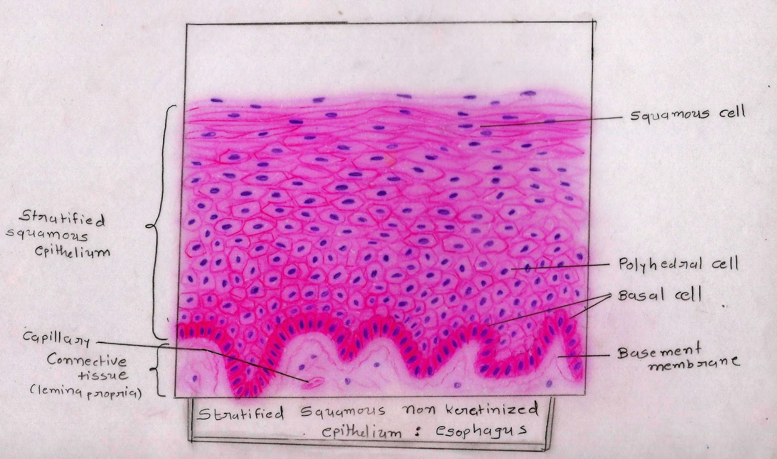
Click the button below to reveal the answer key: They have no nucleus or organelles. A stratified squamous epithelium consists of squamous epithelial cells arranged in layers upon a basal membrane.
Stratified Squamous Epithelium Under Microscope 400x
They are filled with a protein called keratin, which is what makes our skin waterproof.
(2) lining of esophagus, (3) lining distal portion of urethra;
The epithelium at these locations do not have thick layers. Although this epithelium is referred to as squamous, many cells within the layers may not be flattened; Underlying cell layers can be made of cuboidal or columnar cells as well. This is an epithelial layer that consists of many layers of cells.
Nonkeratinized epithelium is a stratified squamous epithelium found in lips, buccal mucosa, alveolar mucosa, soft palate, the underside of the tongue, and floor of the mouth.
Not only are they flat, but they are no longer alive. Cells are gradually pushed toward the surface by the production of newer cells. For the sse label, try to bracket off a region of that type of epithelium, rather than just pointing to 1 cell. The deeper layers are polyhedral (asterisks), and the basal layer is cuboidal (arrows).
The other layers adhere to one another to maintain structural integrity.
Along with a key that defines what each label in (n= nucleus, etc.). Only one layer is in contact with the basement membrane; Stratified epithelium is classified by the cell type on the uppermost layer. Simple squamous epithelium 400x recommended simple cuboidal epithelium 400x recommended draw from side and from on high label nuclei where.
The epithelium at these locations have varying thickness of kertain on top, and thus.
This epithelium shows a basal. Stratified epithelium is classified by the cell type on the uppermost layer. Notice that is true for the pictures shown below. / stratified squamous epithelium, 400x / stratified squamous epithelium;
A stratified squamous epithelium is a tissue formed from multiple layers of cells resting on a basement membrane, with the superficial layer(s) consisting of squamous cells.
(5) lining lower 1/3 of anus. It can be found in the lip, tongue, vagina, anal canal, esophagus, and palate(as in this case, soft palate specifically). A stratified squamous epithelium is made up of a number of layers and the cells of the outer layers are flat (squamous). Unlike keratinized epithelium, nonkeratinized epithelium is moist, and it contains living cells in the surface layer.
Notice that is true for the pictures shown below.
This epithelium covers internal body surfaces which are exposed to some degree of physical trauma. They are typically found in locations where constant abrasion is likely, such as mouth, esophagus and vagina. It is found in moist cavities subject to trauma, whereas the keratinized type is found on dry. Body surfaces internal cavities and tubes parenchyma of glands:
They are typically found in locations where constant abrasion is likely, such as skin.
Fest арна membrane 40% tis (b) nonkeratinized stratified squamous opithelium (a) survey photomicrograph mark nielsen mas new boox 400x (c) sectional view nonkeratinized stratified squamous epithelium 1 • connective tissue • nucleus of epithelial cell in basal layer of epithelium • nucleus of squamous epithelial cell • stratified. The boxed area is presented at a higher magnification in the next slide. Stratified squamous epithelium view related images. The labels are l for lumen, ct for connective tissue, n for nucleus, and sse for simple squamous epithelial cell.
It is named for the shape of the cells on the surface of the tissue.
Stratified squamous keratinized epithelium 400x (palmar skin) the cells on the surface of stratified squamous keratinized epithelium are very flat. 200x note that the superficial layer of cells are flatted (arrowhead) and not cornified (keratinized). Key facts about the stratified epithelium; If you need to draw a tissue at two magnifications or from two different vantages, you can split the field of view (circle) and draw one on one side of the line and the other on the other side:
Simple squamous epithelial 20x labeled.
A picture of simple squamous epithelial cells from a human artery and vein slide. It protects the tissue from damage and water loss. This is due to the convention of. The cells in this tissue are not all squamous (flat).
Nonkeratinized epithelium is a stratified squamous epithelium which lines the buccal cavity.
Cells in the deep layers are basically cuboidal but become more and. This epithelium shows a basal layer of cuboidal cells, then several layers of polygonal cells that become progressively more flat, until they become squamous at the luminal surface. In the deepest layer new cells are produced by the division of stem cells.


