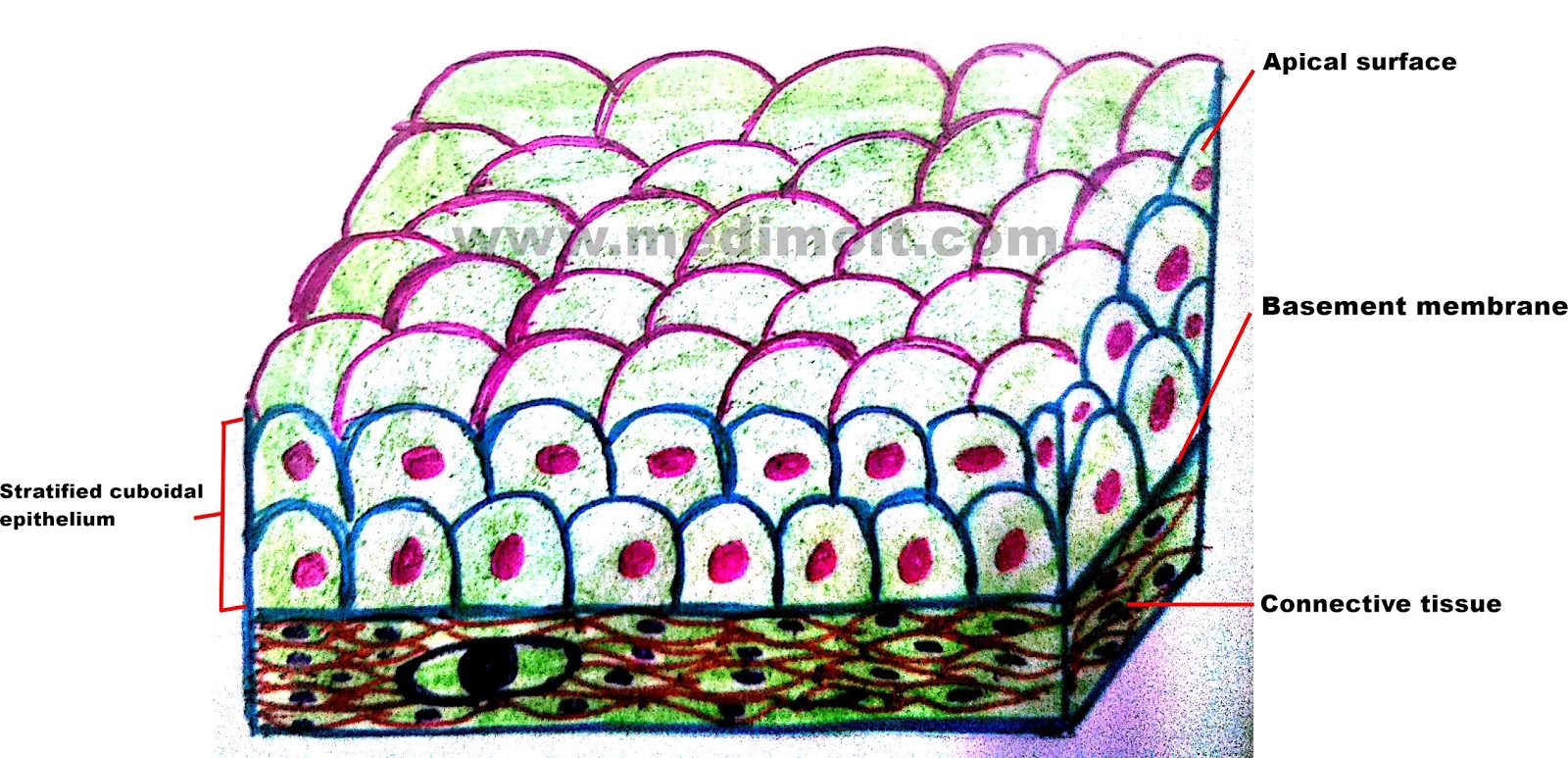
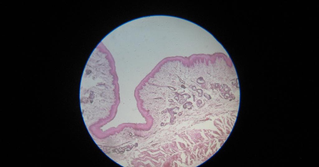
This type of tissue can either be keratinized, or nonkeratinized. Vagina, cervix and oral cavity. Stratified squamous epithelium is composed of multiple layers of flat cells.
Stratified Squamous Epithelium Definition and Function
In fact, this specific role is.
Download scientific diagram | the nonkeratinized stratified squamous epithelium of the tongue dorsum displaying the str.
We identified it from trustworthy source. It is named for the shape of the cells on the surface of the tissue. Efficient monte carlo modelling of individual tumour cell. A typical example of stratified squamous keratinized epithelium is the epidermis.
The cells in this tissue are not all squamous (flat).
Sets found in the same folder. Layers at the surface where keratin cements the debris of dead squamous cells, the non living keratin layer. They are typically found in locations where constant abrasion is likely, such as mouth, esophagus and vagina. Learn vocabulary, terms, and more with flashcards, games, and other study tools.
The keratinization, or lack thereof, of the apical surface domains of the cells.
(2) lining of esophagus, (3) lining distal portion of urethra; Here are a number of highest rated stratified squamous non keratin epithelium pictures upon internet. As the most important difference between the simple epithelium and the stratified epithelium is the number of the layer of cells, the functions of these layers also. These surfaces must be kept moist by bodily secretions to prevent them from drying out.
The nonkeratinized or moist, tissue contains living cells in the inner and outermost layers.
The epithelium at these locations do not have thick layers. Notice that is true for the pictures shown below. Are branched forms of microvilli. Cytoplasmic ratio and hyperchromatic nuclei.
Learn vocabulary, terms, and more with flashcards, games, and other study tools.
Its submitted by direction in the best field. So, the correct option is 'vagina, cervix and oral cavity'. A layer of fluid covers the outer cell layers, and thus providing a moist environment. It is a type of epithelium in which the cells do not contain a huge amount of keratin, but rather are moisturized by mucus from the salivary or the mucus glands.
Layer where cells are continually dividing by mitosis.
Stratified epithelium is classified by the cell type on the uppermost layer. I will show you everything about the rumen histology slide with their identification point and labeled diagram. Download scientific diagram | transversal sections of the esophagus. Is anchored on its basal surface to a basement membrane e.
Stratified squamous non keratin epithelium.
Is adapted to withstand abrasive forces c. The stratified squamous epithelium consists of several layers of cells, where the cells in the apical layer and several layers present deep to it are squamous, but the cells in deeper layers vary from cuboidal to columnar. The function of stratified epithelium is mainly protection. Basale (sb) with melanocytes ( * ).
Terms in this set (4) squamous epithelial cell.
The flattened cells of the surface layer maintain their nuclei and most metabolic functions. (5) lining lower 1/3 of anus. Form a brush border in absorptive cells. The arrow indicates one of these squamous cells.
Layer that is exposed to the outside of the cell.
Click the button below to reveal the answer key:


:watermark(/images/watermark_only.png,0,0,0):watermark(/images/logo_url.png,-10,-10,0):format(jpeg)/images/anatomy_term/stratified-squamous-non-keratinized-epithelium-6/nRrdi4AvLvJbGIysjEvrsA_Stratified_Squamous_non-keratinized_epithelium.png)


