The masticator space is one of the deep compartments of the head and neck. Tumor invasion of the masticator space usually upstages the original tumors. Tumor invasion of the masticator space usually upstages the original tumors.
Masticator Space, Buccal Space, and Infratemporal Fossa
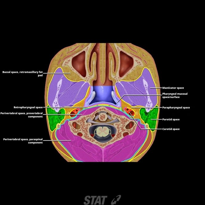
The masticator space is a distinct deep facial space, bounded by the superficial layer of deep cervical fascia and containing the four muscles of mastication and the ramus and posterior body of the mandible.
The masticator space is a fascial space that can be invaded by tumors from adjacent structures or from hematogenous metastases.
Familiarity with the anatomy of the masticator space and its anatomic relationship with. The masticator space lays posterior to the buccal space, anterior to the parotid space, anterolateral to the parapharyngeal space, superior to the. The masticator space is a deep facial space with a complex anatomical structure. In this article, we review the ct and mri features of secondary involvement of the masticator space in a variety of tumors.
The masticator space is a deep facial space that is outlined by the superficial layer of the deep cervical fascia , , and lies laterally and evenly in front of the prestyloid space, medial to the pharyngeal space and beneath the skull base.
It is located between the buccinator. The masticator space as an anatomical and functional entity centered on the mandibular ramus, which divides. The masticator space is a deep facial space with a complex anatomical structure. Anatomy of the masticator space.
The anatomy of the masticator space and its anatomic relationship with adjacent structures is important for imaging interpretation.
Familiarity with the anatomy of the masticator space and its anatomic relationship with. These two layersfusealong the anterior and posterior borders of the mandibular ramus, enveloping the space [2, 3](fig.1). Trismus often complicates evaluation of masticator space disease. Secondary masticator space involvement is not rare.
Normal anatomy and abnormalities the buccal space is an anatomical compartment lying anterior to the masticator space and lateral to the buccinator muscle.
Primary tumors are uncommon, usually benign and of a vascular or neural origin. Ct and mr imaging of the buccal space: The masticator space is situated laterally to the medial pterygoid fascia and medially to the masseter muscle. Each space is delineated by a superficial layer of the deep cervical fascia (sldcf).
The masticator space is a deep facial space that is outlined by the superficial layer of the deep cervical fascia and lies laterally and evenly in front of the prestyloid space, medial to the pharyngeal space and beneath the skull base.
In this article, we review the ct and mri features of secondary involvement of the masticator space in a variety of tumors. Since the major purpose of imaging is to define the likely anatomic origin and also the extent of a given lesion, thorough The purpose of the present study was to precisely define the masticator space to eliminate the use of obsolete and confusing terms to describe the area, and to illustrate the common mass syndromes. Gross anatomy the masticator space are paired suprahyoid cervical spaces on each side of the face.
The purpose of the present study was to precisely define the masticator space to eliminate the use of obsolete and confusing terms to describe the area, and to illustrate the common mass syndromes.
We focus on showing various patterns of tumor spread to the masticator space. The secondary tumor may also extend intracranially from the masticator space along the neurovascular bundle. Secondary masticator space involvement is not rare. The masticator space (ms) is a deep facial space which contains the mandibular ramus, muscles of mastication and the mandibular branch (v3) of the trigeminal nerve.
The buccal space, also known as the buccinator space, is one of the seven suprahyoid deep compartments of the head and neck.
The masticator space contains the mastication muscles, posterior mandible, and mandibular nerve [3, 4]. The purpose of the present study was to precisely define the masticator space to eliminate the use of obsolete and confusing terms to describe the area, and to illustrate the common mass syndromes. Primary tumors are uncommon, usually benign and of a vascular or neural origin. Primary tumors are uncommon, usually benign and of a vascular or neural origin.
The ct and mri features of secondary involvement of the masticator space in a variety of tumors are reviewed to show various patterns of tumor spread to the masticsator space.
Each space is enveloped by the superficial (investing) layer of the d. It may be affected by developmental, neoplastic or infectious lesions. We focus on showing various patterns of tumor spread to the masticator space. Anatomy of the masticator space.
Figure 1 shows that the four mastication muscles are the medial and lateralpterygoids,masseter,andtemporalis[3,4].thelateral pterygoid muscle,.
The masticator space is a deep facial space with a complex anatomical structure. A axial and b coronal t1w mr images show the superficial layer of the deep cervical fascia ( white line) enveloping the space, the ramus of the mandible (r), masseteric muscle (m), medial pterygoid muscle (mp), lateral pterygoid muscle (lp), and temporalis muscle (t).