Type 1 maps to band 14q11.2 and is caused by mutations in the gene for keratinocyte transglutaminase 1, an enzyme responsible for the assembly of the keratinized envelope. With optimal supportive care, the thickened stratum corneum usually resolves in 2 to 4 weeks but can persist, especially in infants with lamellar ichthyosis. Localised to scalp and trunk (warmer sites of the body)
Medicine by Sfakianakis G. Alexandros Skin disease in
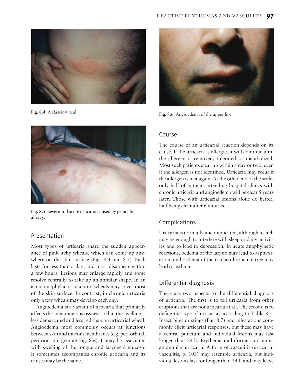
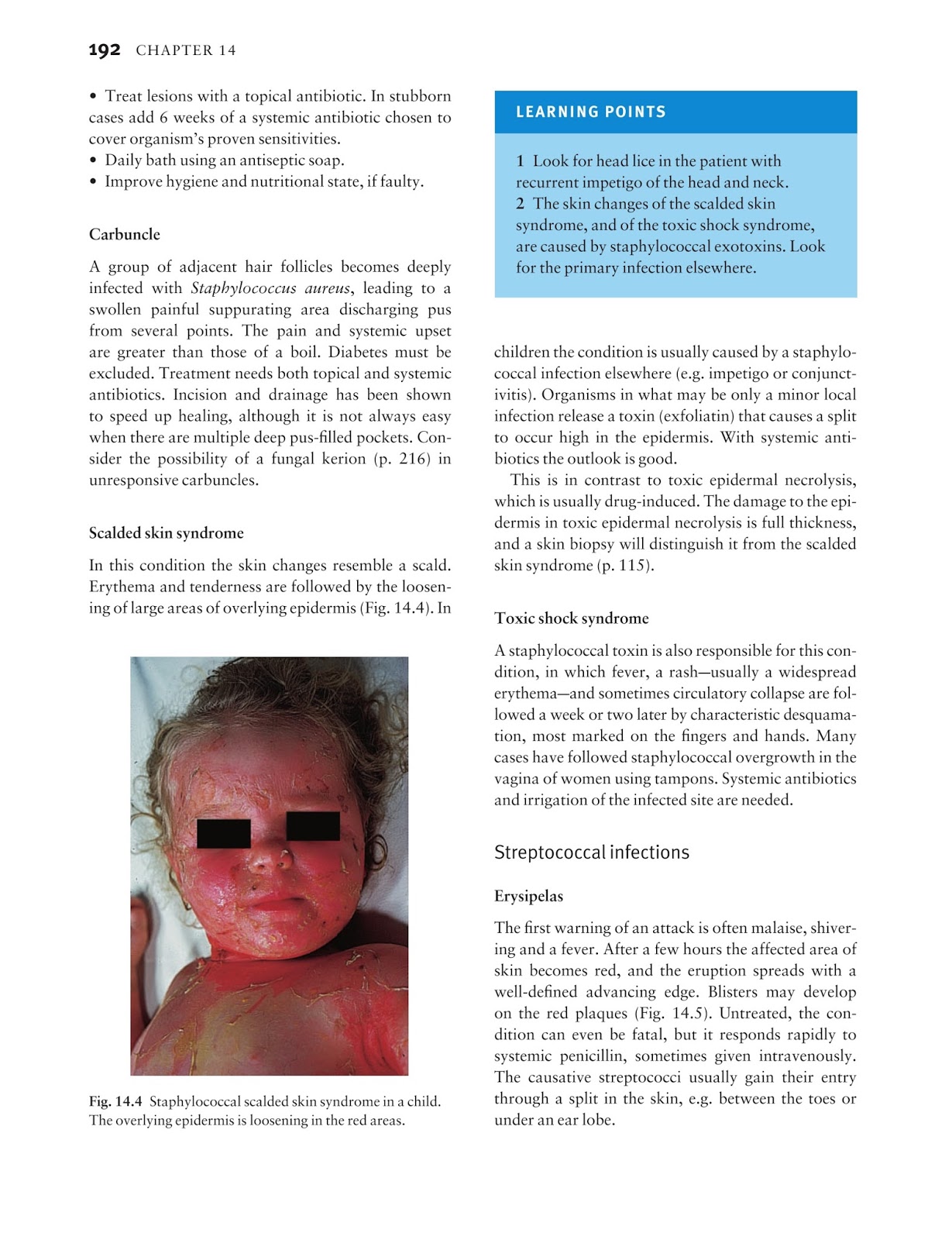
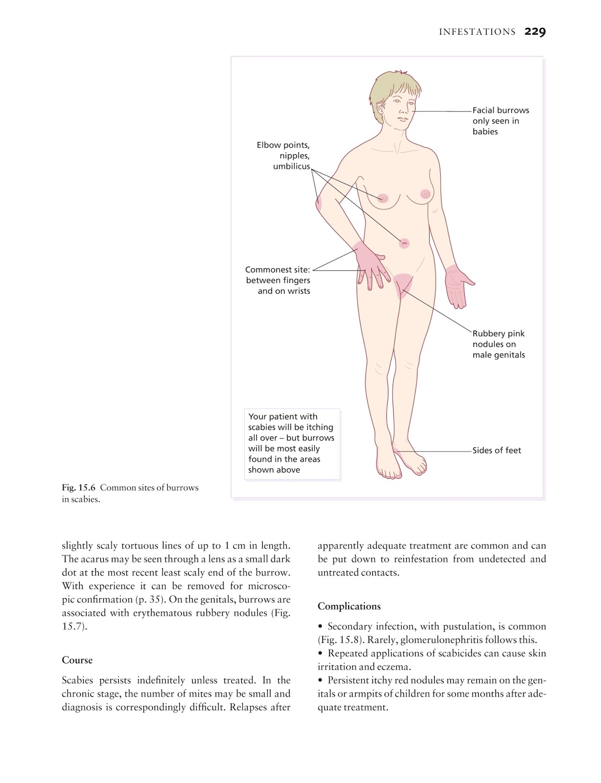
Lamellar ichthyosis is a condition that mainly affects the skin.
These pores and skin abnormalities have an effect on the form of the eyelids, nostril, mouth, and ears, and restrict motion of the legs and arms.
Only when a person receives two recessive genes for lamellar ichthyosis or two for congenital ichthyosiform erythroderma or two for harlequin ichthyosis (etc.) will he manifest one of these disorders. There are some types of ichthyosis, however, where there are associated findings in organ systems other than the skin. Lamellar ichthyosis, also known as ichthyosis lamellaris and nonbullous congenital ichthyosis, is a rare inherited skin disorder, affecting around 1 in 600,000 people. Although most neonates with autosomal recessive congenital ichthyosis (arci) are collodion babies, the clinical presentation and severity of arci may vary significantly, ranging from harlequin ichthyosis, the most severe and often fatal form, to lamellar ichthyosis (li) and (nonbullous) congenital ichthyosiform erythroderma (cie) in older individuals.
Deficiency in steroid sulfatase, which removes proadhesive cholesterol sulfate from intracellular spaces clinical features.
They are caused by recessive genes which ordinarily are dominated by genes for normal skin. The neonate is encased in an “armor” of thick scale plates separated by deep fissures. Infants are usually born encased in a collodion membrane which sheds within a few weeks. Surgical release of constrictive plaques has been previously demonstrated, but the perioperative and intraoperative considerations surrounding this infrequent inte.
It is a less severe phenotype when compared with harlequin ichthyosis (described below), yet still has significant associated morbidity and mortality.
The other two are known as harlequin ichthyosis and congenital ichthyosiform erythroderma. The tight skin around the eyes and mouth often leads to ectropion (outturning of the eyelids) and. 25 rows lamellar ichthyosis is a rare genetic condition that affects the skin. Harlequin ichthyosis is a rare congenital abnormality with more striking appearance and a graver prognosis than for collodion baby.
Mutations in the abca12 gene cause harlequin ichthyosis.
The lamellae form the skin's hydrophobic sphingolipid seal. There is bilateral ectropion and eclabium, and the nose and ears are flattened and appear rudimentary. Symptoms of the following disorder may be similar to harlequin ichthyosis. The collodion baby is no clinical entity, but a symptom occurring in several forms of ichthyosis.
Harlequin ichthyosis (hi) (omim 242500) is a rare, severe form of congenital ichthyosis, which may be fatal.
Infants affected by lamellar ichthyosis are generally born with a shiny, waxy layer of skin (called a collodian membrane) that is typically shed within the first two weeks of life. Harlequin ichthyosis is a extreme genetic dysfunction that primarily impacts the pores and skin. Harlequin ichthyosis is a severe genetic disorder that mainly affects the skin. All arci conditions are considered a clinical spectrum.
Paller will test, secukinumab, has already been highly effective in psoriasis, a more common skin disorder with an increase in this th17 pathway, leading to inflammation and scaling.
In ichthyosis, the skin barrier is abnormal, so the skin is inflamed, dry and scaly. A chronic, congenital ichthyosis inherited as an autosomal recessive trait. Associated findings most forms of ichthyosis have clinical findings limited to the skin. The newborn infant is covered with plates of thick skin that crack and split apart.
In some cases, scales are so thick that they resemble armored.
Lamellar ichthyosis is a rare, autosomal recessive, genetically heterogeneous skin disease caused by mutations involving multiple genetic loci. Lamellar ichthyosis may also cause reddened skin (erythroderma), thickened skin on the palms and soles and decreased sweating with heat intolerance. The extruded lipids are arranged into lamellae in the intercellular space with the help of concomitantly released hydrolytic enzymes. These findings are thought to be caused by the same genetic abnormality that is responsible for the ichthyosis.
Restricted movement of the chest can lead to breathing difficulties.
The thick plates can pull at and distort facial features and can restrict breathing and eating. Ichthyosis lamellaris has an autosomal recessive pattern of inheritance. The collodion baby is less severely affected than the infant with harlequin ichthyosis and may represent a phenotypic expression of several genotypes. Infants with this condition are typically born with a tight, clear sheath covering their skin called a collodion membrane.
The skin beneath the collodian membrane is red and scaly.
It is one of three genetic skin disorders called autosomal recessive congenital ichthyoses (arci). These affect the shape of the eyelids, nose, mouth, and ears and limit movement of the arms and legs. This membrane usually dries and peels off during the first few weeks of life, and then it becomes obvious that affected babies have scaly skin, and eyelids. In our patient the collodion skin was a symptom of ichthyosis congaenita (lamellar ichthyosis).
Lamellar ichthyosis (li) is a rare genetic skin disorder that is present at birth.


