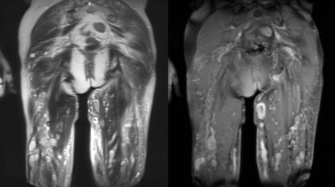
Klippel trenaunay syndrome (kts) is a vascular malformation syndrome comprising of varying involvement of cutaneous capillaries, veins, and lymphatics with hypertrophy of soft tissue and bones of the affected limb. In 1900, the french physicians klippel and trénaunay (, 1) first described a syndrome characterized by a capillary nevus of the affected extremity, lateral limb hypertrophy, and varicose veins. It is imperative that both the radiologist and surgeon be aware of this entity, as.
KlippelTrénaunayWeber syndrome Radiology Case
Klippel and trenaunay first described a syndrome characterized by a capillary nevus of the affected extremity, lateral limb hypertrophy, and varicose veins.
The exact pathophysiology and genetic etiology of the disorder are unknown.
Possible additional features include 14): These patients are at risk for sequestration of platelets, resulting in kasabach. It is imperative that both the radiologist and surgeon be aware of this entity, as. The prevalence of ktws is 1 :
Functionally important arteriovenous fistulas must be approached surgically but ligation and stripping of varicose veins often leads to severe vascular problems with edema and.
Most cases involve the lower limb as the site of malformations. This syndrome has three characteristic features: Prenatal diagnosis using ultrasound has been reported. Weber noted the association of these findings with arteriovenous malformation in the 1900s.
3d us may reveal leg width difference.
Bliznak and staple suggested intrauterine damage to the sympathetic ganglia or intermediolateral tract leading to dilated microscopic arteriovenous anastomoses as the cause. Capillary malformations, soft tissue or bone hypertrophy, and varices or venous malformations. In 1918, weber ( , 2 ) noted the association of these findings with arteriovenous fistulas.


