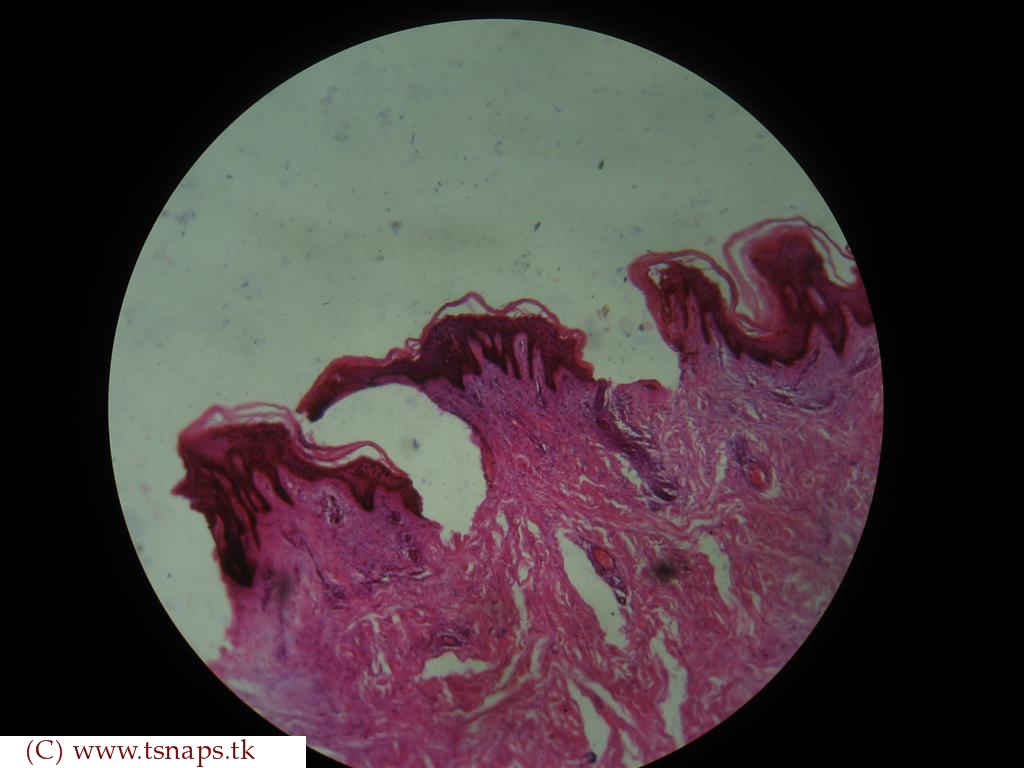
They are filled with a protein called keratin, which is what makes our skin waterproof. As these cells differentiate they are pushed toward the surface by new cells in the basal layer. Slide 43 skin, sole of foot.
Stratified Squamous Epithelium Keratinized cloudshareinfo
Stratified squamous epithelium (keratinizing and nonkeratinizing):
Thick skin is covered by a stratified squamous keratinized epithelium.
The keratin layer is much thicker than the cellular layer. Thin skin is covered by a stratified squamous keratinized epithelium. This specimen has a well preserved epithelium with excellent cellular definition. Label the structures on the slide of keratinized stratified squamous epithelium.
Note the flattened, nucleated surface cells, the “middle zone” of the polyhedral shaped cells, and the basal layer of the polyhedral shaped cells, and the basal layer of columnar cells which rests on.
Stratified squamous keratinized epithelium 400x (palmar skin) the cells on the surface of stratified squamous keratinized epithelium are very flat. Slide 115 , which you used to study bone and the respiratory system, is a longitudinal section through the palate and includes the lip, gingiva, hard palate, and a portion of the soft palate [orientation]. Thus, the outermost layer is still cellular and contains a nucleus. • label it with the following terms:
It is a multilayered epithelium that mainly protects against abrasion and dehydration.
Stratum corneum stratum basale stratum spinosum stratum granulosum dermis reset zoom. Stratified epithelia are classified by the shape of the surface layer of cells. Stratified squamous epithelium covers the tongue, skin and several other organs. Notice the flattened nuclei of the surface layer cells here, causing this to be classified as stratified squamous epithelium.
Layer c is the stratum granulosum, and the basophilic structures in the cytoplasm is.
Label the structures on the slide of keratinized stratified squamous epithelium. Use slides 1 & 2 from the chapter 4 slides for inspiration. The oral mucosa varies from site to site within the oral cavity, but everywhere the epithelium is protective stratified squamousstratified squamous. Epithelial cell renewal the stratified squamous epithelium of skin is replaced in approximately 28 days.
The lining of the human mouth is a standard example of stratified squamous epithelium.
Ultimately, the cells become keratinized and slough off. Anatomy and physiology questions and answers. This type of epithelium is found in mucous membranes. The basal layer is the stratum basale (a), the next several layers of cells is the stratum spinosum (b).
Keratinized stratified squamous epithelium is a type of stratified epithelium that contains numerous layers of squamous cells, called keratinocytes, in which the superficial layer of cells is keratinized.
The flat cells shown on the slide form several layers that make up this type of epithelial tissue. This is the lining of the esophagus, where it is no longer necessary to have an outer keratinized layer to protect against desiccation, as it was for skin. This type of epithelium comprises the epidermis of the skin. View available hint(s) simple columnar epithelium stratified squamous epithelium keratinized stratified squamous epithelium
Keratinized stratified nuclei epidermis squamous epithelium keratinocytes dermis stratum stratum stratum corneum granulosum spinosum stratum basale free space ;
The epithelium of the esophagus is composed of which type of epithelial tissue? The outermost keratinized layer is composed It is found in moist cavities subject to trauma, whereas the keratinized type is found on dry surfaces. It can be found in the lip, tongue, vagina, anal canal, esophagus, and palate(as in this case, soft palate specifically).
Stratified squamous nonkeratinizing epithelium lines the lumen of the esophagus.
The epidermis is keratinized stratified squamous epithelium. Only a few layers of epithelial cells. This epithelium is partially keratinized on gums and hard palate and on filiform papillae of tongue; The epithelial cells form between 10 and 20 layers.
Not only are they flat, but they are no longer alive.
In a section of the tongue, the surface cells and their nuclei are flat (blue arrow) while the deepest cells are columnar or cuboidal (orange arrow). They have no nucleus or organelles. Histology slide courtesy of mt. It protects the tissue from damage and water loss.
You might find out the dermal papillae, sebaceous glands, sweat glands, arrector pili muscles from the skin histology slide.
This epithelium contains 5 layers: You will find the papillary layer and reticular layer in the dermis of the skin. Epithelial tissue covers or lines body surfaces as well as serving to absorb,. • draw a color picture of a keratinized stratified squamous epithelium.
Cells in the stratum basale undergo mitosis to provide for cell renewal.
Use the isolated cheek cells on this prepared slide to illustrate the morphology of typical squamous cells. Stratified squamous epithelium, noncornified/nonkeratinized (moist) on slide 131, esophagus (h&e) identify the noncornified/nonkeratinized, stratified squamous epithelium. A closer examination reveals the outlines of cells within the keratin. The epidermis of the skin is very thin and lines with the keratinized stratified squamous epithelium.


