Stratified squamous epithelium of the esophagus is of the nonkeratinizing type (fig. Keratinized epithelium and nonkeratinized epithelium are two stratified squamous epithelia. Not only are they flat, but they are no longer alive.
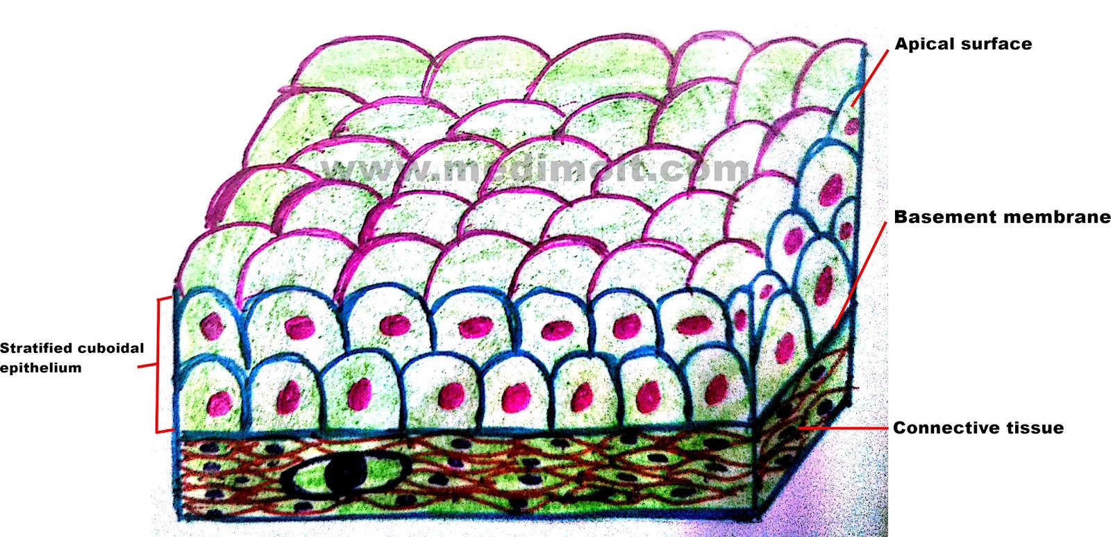
Histology Image Membranous epithelium
Keratinized stratified squamous epithelium eight to twelve circumvallate papillae are located along the sulcus terminalis, separating the anterior from the posterior portion of the tongue.
It is named for the shape of the cells on the surface of the tissue.
The parts of the mouth that feel a little rough such as the upper surface of the tongue and the hard palate at the roof of the mouth contain keratinized epithelia. They have no nucleus or organelles. Simple cuboidal epithelium total magnification: Key facts around the forms of epithelium.
Epithelium wikipedia from upload.wikimedia.org this illustration is included in.
This type of epithelium comprises the epidermis of the skin. Notice the flattened nuclei of the surface layer cells here, causing this to be classified as stratified squamous epithelium. Use slides 1 & 2 from the chapter 4 slides for inspiration. Laboratory exercise 7 epithelial tissue instructions:
A keratinized stratified squamous epithelium consists of basal spinous and cornified layers from the basement membrane to the surface appendix fig.
They have no nucleus or organelles. Keratinized stratified squamous epithelium is a type of stratified epithelium that contains numerous layers of squamous cells, called keratinocytes, in which the superficial layer of cells is keratinized. The arrow indicates one of these squamous cells. The oral cavity is lined by a mucous membrane (the oral mucosa) consisting of a stratified squamous epithelium, which may or may not be keratinized,.
Stratified squamous keratinized epithelium 400x palmar skin the cells on the surface of stratified squamous keratinized epithelium are very flat.
This nonkeratinized epithelium is mostly moist and acts as a less effective barrier. Surface cells are alive and kept moist. Although stratified squamous keratinized epithelium covers the entire surface of the body,. Multiple layers of flattened cells, basal cells typically are cuboidal, while apical cells are squamous.
· in the stratified squamous epithelium, only one layer is directly attached.
These tissues are formed by four layers: As the cells move upwards, they accumulate keratin in the process of keratinization, where they become thin, metabolically inactive pockets (squames) of keratin lacking nuclei. This epithelium contains 5 layers: Draw the microscopic features of the indicated structure or organ using colored pencil 2.
Tap card to see definition 👆.
On the basis of the cytoskeletal structures seen within each cell, these tissues may be divided into two categories: The stratified squamous epithelial cells that lack the keratin protein are called the nonkeratinized epithelium. Keratinized tissues are particularly significant in situations where there is physical abrasion as well as the likelihood of desiccation and water. The types of epithelium are the following:
Stratified epithelia are classified by the shape of the surface layer of cells.
This epithelium tends to be strong and resistant to mechanical stress. A stratified squamous keratinized epithelium is found on surfaces subject to the abrasion that occurs with mastication, e.g., the roof of the mouth (palate) and gums (gingiva). The cells in this tissue are not all squamous (flat). Nonkeratinized epithelium is a stratified squamous epithelium which lines the buccal cavity.
Draw the schematic picture of each type of connective tissue.
Stratified squamous keratinized epithelium 400x (palmar skin) the cells on the surface of stratified squamous keratinized epithelium are very flat. There are several different types of stratified squamous epithelia. Keratinized epithelium is a stratified squamous epithelium that forms the epidermis of land vertebrates. Keratinized stratified nuclei epidermis squamous epithelium keratinocytes dermis stratum stratum stratum corneum granulosum spinosum stratum basale free space
• draw a color picture of a keratinized stratified squamous epithelium from thick skin.
Slide 115 , which you used to study bone and the respiratory system, is a longitudinal section through the palate and includes the lip, gingiva, hard palate, and a portion of the soft palate [orientation]. The basal layer, the spinous layer, the. Label the parts indicated on the checklist. This consists of two to three layers of rounded basal cells in the basal region that are small with a high nuclear :
Squamouscuboidal (with microvili, without microvili) columnar (with microvili, via surface cilia, via stereocilia, with pseudostratification) stratified epithelium.
Stratified squamous epithelium (keratinized) specimen: They are filled with a protein called keratin, which is what makes our skin waterproof. Click card to see definition 👆. Stratified squamous keratinized epithelium 40x (palmar skin).
Keratinized stratified squamous epithelium keratin is a tough, fibrous intracellular protein that helps protect skin and underlying tissues from heat, microbes, and chemicals.
Each papilla is surrounded by a deep sulcus that receives ducts of the serous glands of von ebner.