Horseshoe kidney is commonly seen as part of other anomalies such as trisomy 18 (25%), caudal dysplasia syndrome, and zellweger syndrome. Children who have horseshoe kidney have one “fused” kidney instead of 2 separate kidneys. Horseshoe kidney is a medical condition in which 2 kidneys are fused together and resemble a horseshoe in appearance.
Horseshoe Kidney Photograph by Medimage/science Photo Library
By fusing, they form a u shape, which gives it the name horseshoe. horseshoe kidney occurs during fetal development, as the kidneys move into their normal position in the flank area (area around the side, just above the waist).
They form a u shape like a horseshoe.
Horseshoe kidney is when the two kidneys join (fuse) together at the bottom. However, it's not just the shape and structure of the kidneys that is abnormal. In total, 146 patients with hsk (age of ≥20 years) from two tertiary hospitals were included in this study. Horseshoe kidney is a condition in which the kidneys fuse (bind) together at the bottom, forming a “u” shape or horseshoe shape.
The horseshoe kidney is more common in connection with certain genetic diseases:
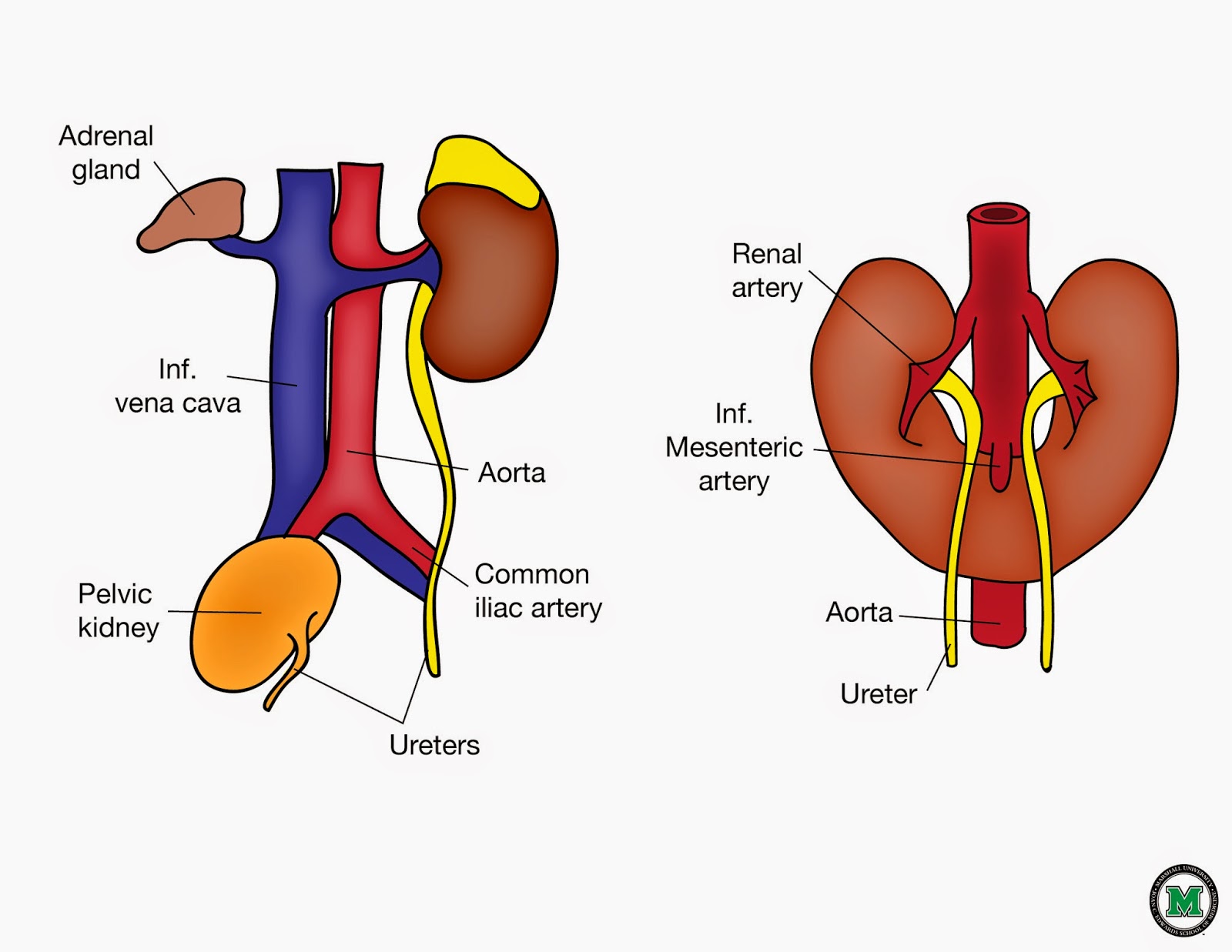
What is a horseshoe kidney? In a crossed renal ectopia, both kidneys are positioned on the same side of the body with one ureter crossing the midline to drain into the bladder while in a fused pelvic kidney there is a single renal mass that is drained by. Horseshoe kidney occurs in about one in 500 children. A horseshoe kidney is formed by fusion across the midline of two distinct functioning kidneys, one on each side of the midline.
They form a shape like a horseshoe.
Horseshoe kidney when the 2 kidneys join (fuse) together at the bottom to form a u shape like a horseshoe. Dysmorphic features include plagiocephaly, malar hypoplasia, broad nasal bridge, poorly developed philtrum and nasal alae, cleft palate and hypodontia. It’s located lower in the pelvis and closer to the front of your body. It occurs during fetal development as the kidneys move into their normal position in the flank area (area around the side, just above the waist).
A horseshoe kidney is also in a different location compared to two typical kidneys.
This is where the disease gets its name. Horseshoe kidney, also called renal fusion, is a condition that starts before a child is born. With horseshoe kidney, as the kidneys of the fetus rise from the pelvic area, they become attached (“fuse”) together at the lower end or base. Horseshoe kidney is a congenital condition, which means it happens before birth while the baby is still developing inside the mother’s womb.
With horseshoe kidney, however, as the kidneys of the fetus rise from the pelvic area, they fuse together at the lower end or base.
It is also known as renal fusion. They are connected by an isthmus of either functioning renal parenchyma or fibrous tissue. The horseshoe kidney is one form of renal fusion abnormality. Horseshoe kidney can occur alone or with other disorders.
A horseshoe kidney is a congenital malformation in which the kidneys don't separate during development.
The kidneys are normally located in the retroperitoneum between the transverse processes of t12 and l3 with the left kidney slightly more superior than the right 6). The horseshoe kidney lies a little deeper on the lumbar spine than normal and is also “the wrong way”, which is why the renal pelvis faces forward towards the abdomen. Horseshoe kidney is a condition in which the kidneys are fused together at the lower end or base. In the vast majority of cases, the fusion is between the lower poles (90%) 13.
Orphanet is a european reference portal for information on rare diseases and orphan drugs.
67 the fusion is typically at the lower poles but can vary greatly in the quantity of fused parenchyma. The horseshoe kidney is one form of renal fusion abnormality. 1 their location is abnormal as well. It consists of two distinct functioning kidneys on each side of the midline, connected at the lower poles (or rarely at the upper poles) by an isthmus of functioning renal parenchyma or fibrous tissue that crosses the midline of the body.
Four cases have been reported in the literature in two unrelated families.
The condition occurs when a baby is growing in the womb, as the baby’s kidneys move into place. Medscape reference provides information on this topic. A very rare syndrome with characteristics of intellectual deficit, horseshoe kidney, and congenital heart defects. During fetal development, two kidneys form in the pelvic region.
Horseshoe kidney (hsk) is a congenital disorder that is usually asymptomatic, but that increases the risks of kidney stones and infectious disease.
The attached kidneys form a horseshoe or u shape. Horseshoe kidney, also called renal fusion, is when two kidneys are fused or joined together. You may need to register to view the medical textbook, but registration is free. As the name suggests, a horseshoe kidney is an abnormality where the two kidneys get fused together to form a horseshoe.
Horseshoe kidney occurs in about 1 in 500 children.
A horseshoe kidney consists of two normal functioning kidneys attached by a band of tissue called the isthmus. A horseshoe kidney consists of two normal functioning kidneys attached by a band of tissue called the isthmus. In the remainder, the superior, or both the superior and. Rather than being present in the upper abdomen, underneath the rib cage and next to your spine, a horseshoe kidney is typically.
The condition occurs when a baby is growing in the womb, as the baby’s kidneys move into place.
It is also known as renal fusion. Horseshoe kidney, also known as ren arcuatus (in latin), renal fusion or super kidney, is a congenital disorder affecting about 1 in 500 people that is more common in men, often asymptomatic, and usually diagnosed incidentally. Chromosome 18 is present three times instead of twice. In a crossed renal ectopia, both kidneys are positioned on the same side of the body with one ureter crossing the midline to drain into the bladder while in a fused pelvic kidney there is a single renal mass that is drained by.
By fusing, they form into a u shape, like a horseshoe.
The characteristic horseshoe shape usually occurs because the bottom part of the two kidneys fuse together and form a bridge that we surgeons call the isthmus. The other two main types are crossed fusion renal ectopia and a fused pelvic kidney. It occurs during fetal development as the kidneys move into their normal position. A horseshoe kidney is ectopic and usually situated anterior to the aorta and vena cava.
It's quite rare, affecting 1 in 500 people.
The horseshoe kidney is the most common type of renal fusion anomaly. The other two main types are crossed fusion renal ectopia and a fused pelvic kidney. Horseshoe kidney occurs as a baby develops before birth.


