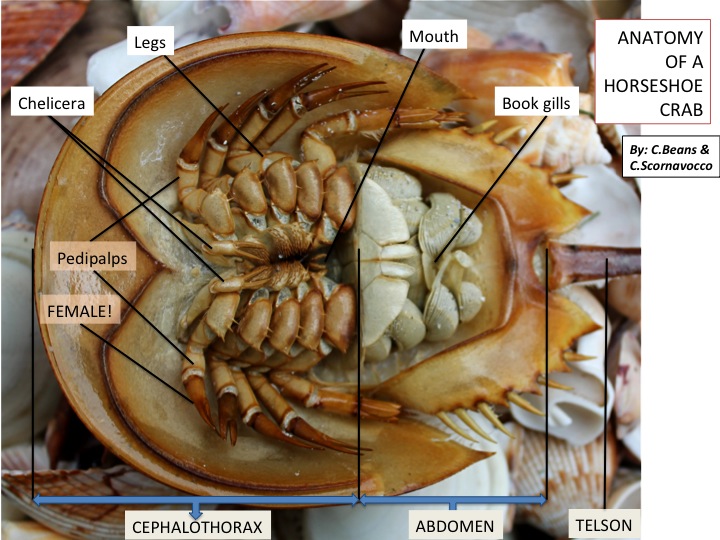
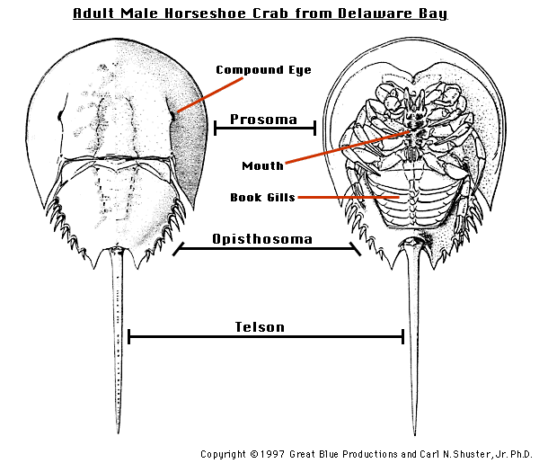
The largest section of the animal, the cephalothorax, houses parts of the intestinal tract. The median eyes have cells sensitive to visible light and others to the ultraviolet range. The anatomy of a horseshoe crab study.
Pin on Animals Wild Life
Migrating birds, such as red knots, are able to eat enough horseshoe crab
(you can mouse over the divisions of the body in the illustration for a closer look) the prosoma contains a sizeable intestinal tract with an esophagus and proventriculus (used to grind food),.
The front section is called the prosoma. They appear at their smallest on the surfaces of the gill leaflets. A distinctive part of a horseshoe crab anatomy is the long, stiff caudal spine. This is an online quiz called horseshoe crab anatomy.
Carmichael, dauphin island sea lab and the university of south alabama, explains the differences and similarities between the male and female hor.
The body of the horseshoe crab is divided into three sections. The prosoma, opisthosoma, and the telson. The horseshoe crabs have book gills, which exchange respiratory gases. You need to get 100% to score the 5 points available.
There are three divisions to the body of the horseshoe crab:
There are three divisions to the body of the horseshoe crab: Inside the crab we find important anatomical features such as the heart, brain, and nervous system. These two legs are called pushers and provide most of. We’ll start with the pair closest to the book gills.
These are sometimes referred to as the cephalothorax, the abdomen, and the tail.
The prosoma , the opisthosoma, and the telson. The middle section is called the opisthosoma. The cephalothorax, abdomen and tail. While they’ve certainly experienced a few evolutionary adaptations, their physiology has remained largely unchanged over time, which is why they’re so often referred to as ‘living fossils.’
The horseshoe crab has many eyes.
These are sometimes referred to as the cephalothorax, the abdomen, and the tail. Up to 24% cash back horseshoe crab anatomy. Two of the secondary eyes are located on the bottom of the animal. Horseshoe crab anatomy the horseshoe crab’s body is divided into three sections.
Three embryonic moltings later (by the 8th day), the legs and book gills are visible.
The prosoma contains a sizeable intestinal tract with an esophagus and proventriculus (used to grind food), a nervous system concentrated into a bulbous brain, a. You must be logged in to post a comment. While this may look dangerous, it is not a weapon and serves only to help the animal in forward movement; While the telson may look dangerous, the
And the horseshoe crab’s tail is called the telson.
As long as the gills are kept. The body of the horseshoe crab is divided into three regions: Posted on july 17, 2017 by wellfleet bay. The carapace of the horseshoe crab is made up of three sections:
You may not know it, but horseshoe crab eggs are very important to birds migrating north along the coast.
Internal anatomy in most undergraduate invertebrate zoology labs the internal anatomy of horseshoe crabs is not studied. The prosoma , the opisthosoma, and the telson. The oldest known horseshoe crab species, (lunataspis aurora) was discovered by scientists in 2008 and is estimated to be nearly 450 million years old. An eye made of many lenses, each with a very limited scope.
The horseshoe crab has two primary compound eyes and seven secondary simple eyes.
The coxa of these legs has a small exopodite (external branch of the appendages) called the flabellum (highlighted in yellow). The horseshoe crab has a hard exoskeleton and 10 legs, which it uses for walking along the seafloor. The fifth set of appendages are well suited to pushing the horseshoe crab across the sandy bottom. There is a printable worksheet available for download here so you can take the quiz with pen and paper.
1288 boston post rd, madison, ct 06443.
Terms in this set (7) cephalothorax. Atlantic horseshoe crabs are one of the many important organisms living in estuaries along the east and gulf coasts of the united states. Most of these are just light receptors though. There are approximately one million sensory cells in this organ alone, which the limulus uses to test the composition of the water.
The nervous system of a horseshoe.
The ventral surfaces of horseshoe crabs, in particular the appendages, are covered in a profusion of setae and stout bristles in a range of sizes (top photograph). The branchial warts occur on the endopodites of the gills and are surrounded by spatulate setae, which sample the exhalant water stream from the gills. When conducting research at wellfleet bay, a lot goes on between the early stages of brainstorming research questions and compiling study results. And to right it, if it gets turned on its back (an important function as none of its limbs reach beyond the edge of the carapace).
The entire body is protected by a hard shell known as a carapace.
The first section is the prosoma, or head. Horseshoe crabs have six pairs, five of them ambulatory. Wet the horseshoe crab can survive on land for a very long time. A body region composed of the head and thorax fused together.
Its head, known as the prosoma, is the largest part of its body and contains most of the nervous systems and vital biological organs including the brain, heart, nervous system, mouth, and several glands.
There are 5 additional eyes on the top of its shell (two median eyes, one endoparietal eye and two rudimentary lateral eyes ). By the fourth day after fertilization, rudimentary appendages can be seen in the egg. The horseshoe crab gets its name from the shape of its head, which is rounded in shape just like the shoe of a horse.


