The full spectrum can only be viewed using a free account. “extra peaks” in a variety of commonly used nmr solvents, in the hope that this will be of assistance to the practicing chemist. 5.8 5.0 5.2 6.1 7.01 experimental sp2 ih chemical shifts (ppm).
1 H NMR spectrum of MAEL. Download Scientific Diagram
This is because the protons from these groups get exchanged with the proton of water or an acid.
Notice that there are three major peaks of differing heights.
1h nuclear magnetic resonance (nmr) spectrum. Br all have one peak in their 1 h nmr spectra because all of the protons in each molecule are equivalent. Nmr spectroscopy is the technique for analysing an organic molecule. This means that in the h nmr the solvent will not give any peaks the same solvent is used in c13 nmr and in this case there will be one peak due to the solvent that will appear on the spectrum.
The two peaks on the spectrum are located at (d)2.42 and (d)1.07.
Provide nmr spectra of your unknown. There are a lot of compounds especially organometallics that give signal at negative ppm, but you will probably. Figure 2 shows the 1 h spectrum of the quinine sample, collected using 8 scans in just over one minute. The area under the peak of a proton signal is proportional to the number of equivalent hydrogen atoms giving rise to the peak.
Sometimes other peaks can be seen around 1 h peaks, known as spinning sidebands and are related to the rate of spin of an nmr tube.
Alkene region modified from earlier handout. In the exam it is likely this peak will not occur on the spectra. One at 1.87 ppm (the equivalent ch 3’s) and the other at 3.86 ppm (the ch 2). The nmr of bromobenzene is shown below.
Nmr signals may have different number of peaks (the number of lines).
The peaks at 2.1 and 0.9 ppm as a ch 2 ch 3 unit. Nuclear magnetic resonance (nmr) spectroscopy. These are experimental artifacts from the spectroscopic analysis itself, not an intrinsic feature of the. A comment about nmr and benzene rings.
What peaks are shown by high resolution h nmr of ethanol?
Splitting and multiplicity (n+1 rule) in nmr spectroscopy. Unless otherwise indicated, all were run at room temperature (24 ( 1 °c). The 1 h nmr spectrum of a compound with one methyl group (ch 3), one methylene (ch 2), and one methine (ch) will have 3 peaks with peak ratios of 3:2:1. Signal splitting is arguably the most unique important feature that makes nmr spectroscopy a comprehensive tool in.
In the above 1 h nmr spectrum of methyl acetate ( fig.
View the full spectrum for free! Ethanol 1 h nmr spectrum (high res.) if the spectrum of ethanol is recorded as a high. Tokyo kasei kogyo company, ltd., tokyo, japan: Assign all possible vibrations on the ir spectrum.
More detailed than the “summary” 90 ii.
A b c d e c d e a b. Ch 3 ha ha hb hb hc As with most organic molecule peak assignment problems, the first step is to collect a 1 h 1d spectrum. However, it is known where this peak is so it can be ignored.
This is the easiest to interpret.
The nmr spectrum of ethyl benzene, c 6 h 5 ch 2 ch 3, is shown below.the frequencies correspond to the absorption of energy by 1 h nuclei, which are protons. Why do nh protons usually appear as broad singlets in h nmr spectra? Download my free ebook 10 secrets to aci. H nmr spectroscopy and interpretation:
Provide ir spectrum of your unknown.
(think of it as the lines you see, l = n + 1, where n = number of equivalent adjacent h) this tells us we that the peaks at 4.4 and 2.8 ppm must be connected as a ch 2 ch 2 unit. What does peak splitting in a proton nmr spectrum tell you? Signal (δ2.46 ppm) appears as two peaks (a. View the full spectrum for free!
At this introductory level, all you can safely say about.
Integration of the area under each peak gives the relative number of hydrogens that peak corresponds to. This is called the splitting of the signal or the multiplicity. The number of peaks correspond to the number of unique, or chemically indistinguishable, hydrogen nuclei. Use the information from nmr spectra and ir experiment and provide a possible structure of your compound.
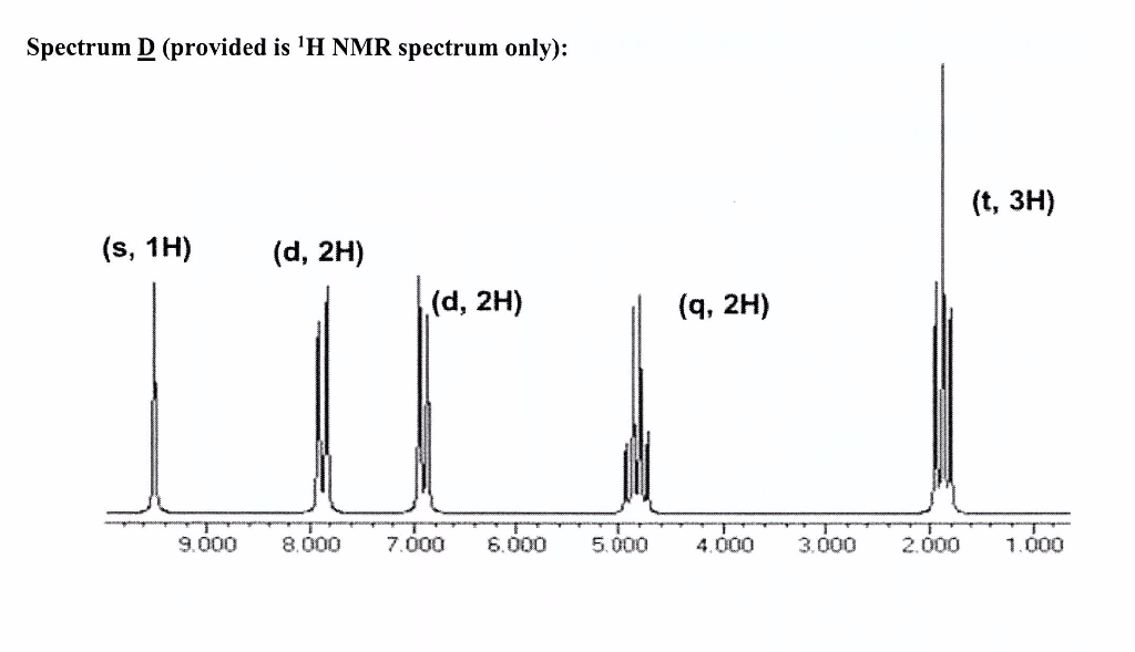
Using the chemical shift charts, the h can be assigned to the peaks as below:
This is a standard reference point with the signal set exactly at 0 ppm and you can ignore it when analyzing an nmr spectrum. The peak at the far right is for the standard reference compound tetramethylsilane (tms, more discussions in chemical shift section 6.6.2 ), not for the compound. Doublet) the ch signal (δ5.84 ppm) appears as four peaks (a. 6.6a ), we can see that there are three signals.
There are two peaks on the graph, therefore of the 10 hydrogens in the molecule, there are two types.
7.2ppm (5h) = arh 4.4ppm (2h) = ch 2 o Nmr is particularly useful in the identification of the positions of hydrogen atoms ( 1 h) in molecules.


