The palatine bones are situated at the back of the nasal cavity between the maxilla and the pterygoid process of the sphenoid bone. Seventy percent of the subjects observed in this. Tensor veli palatini receives arterial blood supply from the greater palatine branch of the maxillary artery, as well as the ascending palatine branch of the facial artery.
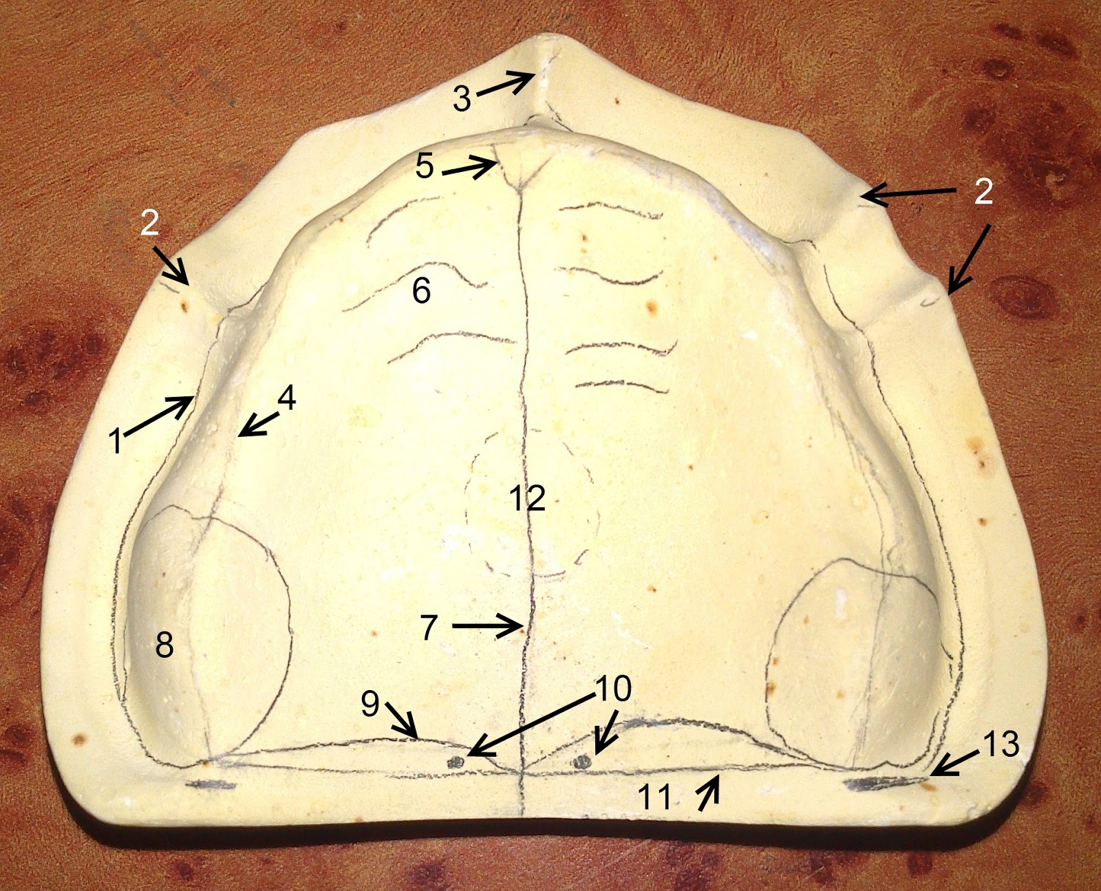
posterior palatal seal
• the fovea are ductal openings into which the ducts of other mucous glands drain.
The floor and lateral walls of the nasal cavity, the roof of the mouth, and the floor of the orbits.
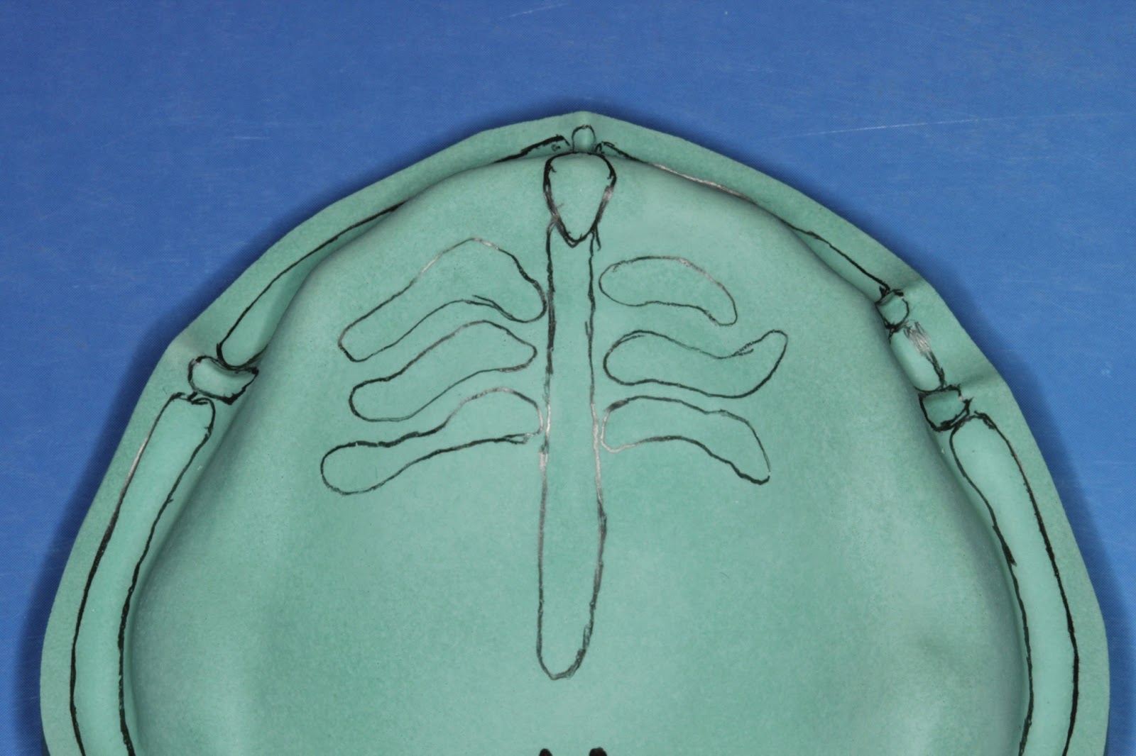
The fovea palatini are two depressions that lie bilateral to the midline of the palate, at the approximate junction between the soft and hard palate and denote the sites of opening of ducts of small mucous glands of the palate ( figure 3 ). The fovea centralis is a small, central pit composed of closely packed cones in the eye. Clinical, radiographic, and histologic studies of the fovea palatini indicate that they were positioned 1.31 mm. Two small depressions in the posterior aspect of the palate, one on each side of the midline, at or near the attachment of the soft palate to the hard palate.
Foveae palatinae, commonly written as fovea palatine or palatine fovea, are two small pits or depressions in the posterior aspect of the palatal mucosa, one on each side of the midline, at or near the attachment of the soft palate to the hard palate.1 they are at border of the maxillary complete dentures.2 they are located in close proximity to the vibrating line and are.
Clinical, radiographic, and histologic studies of the fovea palatini indicate that they were positioned 1.31 mm. • they are the ductal openings into which the ducts of other palatal mucosal glands drain • doesnot represent the junction of hard and soft palate and should be used only as a guideline to placement of posterior palatalseal. The palate of the pigshowsmultiple openings0f the palatal mucous glands. Function bilateral contraction of the tensor veli palatini tenses.
(mean of 100 subjects) in front of the vibrating line.
The fovea palatini are surface landmarks that can be observed clinically in the mouth. One of its functions is to keep the cheeks taut and prevent the cheek from getting in between the occlusal plane. It is located in the center of the macula lutea of the retina. Location in relation to one another, together with the regional innervation and the interrelation of the underlying soft and hard structures.
The fovea is surrounded by the parafovea belt and the perifovea.
The location of the fovea palatini is also important to note in the edentulous patient. They help to form the pterygopalatine and pterygoid fossae, and the inferior orbital fissures. (mean of 100 subjects) in front of the vibrating line. Radiographically and histologically, the foveae were located in soft tissue covering.
The fovea is surrounded by the parafovea belt, and the perifovea outer region.
Radiographically and histologically, the foveae were located in soft tissue covering the hard palate in all specimens. •there are two glandular openings (fovea palatini) within the tissues of the posterior portion of the hard palate, usually lying on either side of the midline. This can be a problem with the older patient whose muscles lose their tonus. Fovea palatini two depressions that lie bilateral to the midline of the palate, at the approximate junction between the soft and hard palate.
Recording posterior palatal seal is an important clinical step thus arbitrarily scraping of the cast using fovea palatini and hamular notch as reference, should be discouraged.
The fovea is responsible for sharp central vision (also called foveal vision), which is necessary in humans for reading, driving, and any activity where visual detail is of primary importance. Clinical, radiographic, and histologic studies of the fovea palatini indicate that they were positioned 1.31 mm. The fovea is responsible for sharp central vision, which is necessary in humans for activities for which visual detail is of primary importance, such as reading and driving. Other pertinent areas of the maxilla are noted on the labeled cast.
Fovea palatini • two glandular openings within the tissues of posterior portion of hard palate, usuallylying on either side of midline.
Permit compression of the tissues. Maintain peripheral seal of the denture. Help overcome poor adaptation of the denture secondary to shrinkage of the acrylic resin during processing. (mean of 100 subjects) in front of the vibrating line.