Dental plaque is made up of different types of bacteria held together in a sticky matrix they create themselves. The microscope and chart were integral to the periodontist’s process. Next, the slide was placed under the microscope and displayed on the screen in the patient’s view.
Bacteria of dental plaque under the microscope YouTube
It is a sticky colorless deposit at first, but when it forms tartar, it is often brown or pale yellow
Over time, dental plaque (a naturally occurring bacterial biofilm) and stains build up on tooth surfaces.
Dental calculus (also called tartar), a harder deposit, can then form both above and below the gum line. How the microscope adds to the joy of being a dental hygienist. Oral and throat cancer screening: In his office, we provided a plaque analysis profile for each patient.
Dr petre chooses to treat these infections in a natural way, so she will make a custom oral care program that fits your needs.
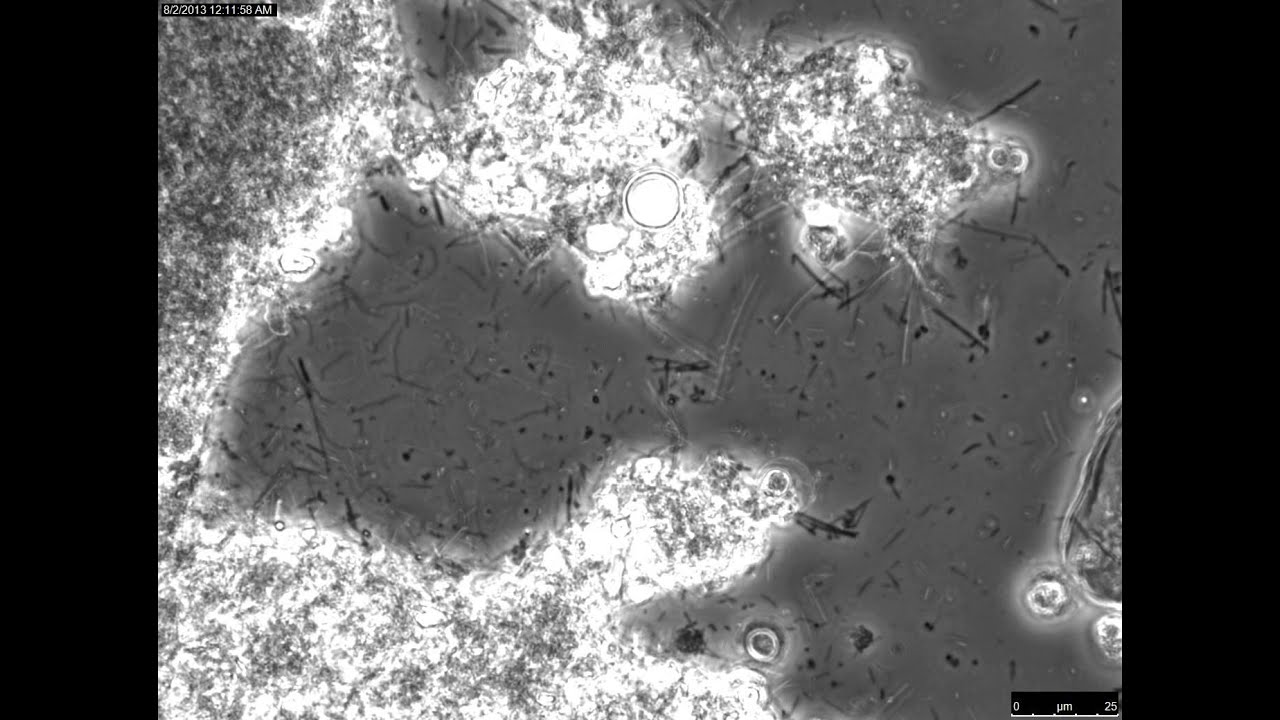
Periodontal disease is a risk factor for heart disease, diabetes, stroke and even pneumonia. Dental plaque, as seen under the microscope. Dental plaque microscopy consists of collecting a small sample of your saliva and scraping off plaque from around some of your teeth. Oral bacteria under light microscope
People with gum disease are statistically more likely to have atherosclerosis, plaque buildup in the arteries.
Leave it on your teeth too long and it can cause some pretty severe damage. Ever experience bleeding, sore, or swollen gums? Microscopic photo of dental plaque. Plaque, that hidden, evil monster in your mouth has a secret.
What plaque looks like under a microscope?what does plaque bacteria look like?dental plaque is a biofilm of microorganisms (mostly bacteria, but also fungi) that grows on surfaces within the mouth.
I tell them that this is how we determine their disease risk. Plaque can also develop under the gums on tooth roots and break down the bones that support teeth. It's easy to see how cavities can form in such a perfect hideout. We use the microscope at your first visit and then every time you come in for teeth cleaning we check your plaque to make sure there is no new infection.
Dental cavities, caused by bacteria, can get under the filling or crown if space has developed and can reinfect the root canal area.
Then the dental plaque is put on a slide under a microscope, connected to a computer monitor to enable you to see what is in your mouth. Run, don’t walk, to your dental office for periodontal treatment if you think you may have symptoms. “i’d like to take a sample of the plaque from under your gums to see if you have the harmful bacteria that cause periodontal infections.” most patients have never heard anything like that before! In fact, most of them are narrower than a single toothbrush bristle but wide enough for bacteria to hide.
Under a microscope, these crevices might look like a deep canyon.
More sticky plaque adheres to this hard irregular tartar surface, and thus it. Well, those are some of the signs that plaque has been resting too long around your teeth and along your gum line. This entailed taking a small sample of the patient’s plaque and saliva and placing it on a slide. This photo shows the dental plaque accumulating on the tooth’s surface.
(dental peeps call it calculus, so if you hear us talk about the “calculus build up”, that’s what we are referring to, no math done here 😉 ) think of tartar as petrified plaque.
This is a look at dental plaque under a microscope: The build up can end up forming something called calculus (also known as tartar) which is a form of hardened dental plaque. Plaque is a sticky film of bacteria that constantly forms on teeth.