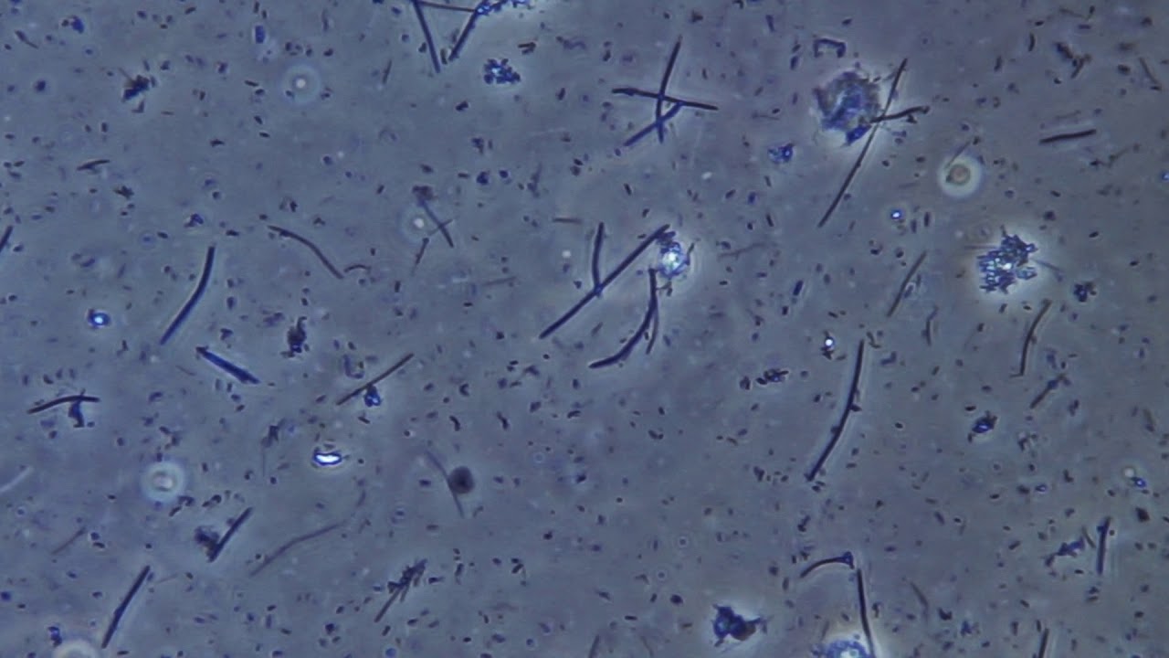
Plaque instability under the microscope. 40x.бактерии зубного налета под микроскопом. Phase contrast of phv, ob.
Scanning electron microscope (SEM) images show extreme
Significant others, restaurants, cooks, dishes, salad bars, buffets, shared food, lipgloss, cigarettes, toothpaste, shared telephones, keyboards, crowded public places, recirculated air in planes, sealed buildings, poorly maintained heating and.
How the microscope adds to the joy of being a dental hygienist.
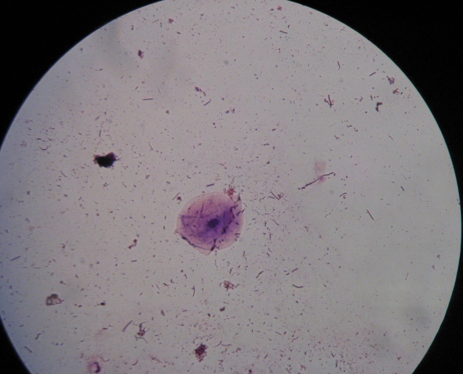
It is a sticky colorless deposit at first, but when it forms tartar, it is often brown or pale yellow We have the actual plaque sample, and attached or surrounding that sample are a few different types of bacteria. Gum health = systemic health. It stains teeth positively revealing where this plaque is, i found the active substance is called erythrosine.
The mean plaque index score of most patients generally decreased during the various treatment phases and hence the overall bacterial count.
“i’d like to take a sample of the plaque from under your gums to see if you have the harmful bacteria that cause periodontal infections.” most patients have never heard anything like that before! Dry samples were directly observed on slides under the microscope at 100× oil objective. What plaque looks like under a microscope?what does plaque bacteria look like?dental plaque is a biofilm of microorganisms (mostly bacteria, but also fungi) that grows on surfaces within the mouth. What we have here is a sample of plaque that we took and mounted on a microscope slide.
Taking a plaque sample and viewing under electron microscope helps to see the health of your gum.
Over the last decade scientists had started to look at ancient plaque samples under a microscope, hoping to find microscopic bits of food stuck in its hard matrix. In this photo we can see bacteria colored on purple and some tiny blood cell in red over the surface of a tooth. Plaque instability under the microscope older newer. “young women had more strokes than young men in a large, united states claims sample”.
Wet calculus samples were mixed with a drop of normal saline (approximately 50 μl) and a coverslip was put on it.
I tell them that this is how we determine their disease risk. The association between poor dental hygiene and incident ischemic stroke; I’ll show you a couple of things here. The preparations were observed under 40× objective.
These were then assessed under a microscope to carry out a quantitative and qualitative assessment of the plaque.
Today these stains are going to be used with microscopy samples to enhance contrast. Bacteria of dental plaque under the microscope. The pictures were taken with a microscope that scans a sample with a focused beam of electrons, and the images are then coloured either digitally or by hand to distinguish the decay and plaque and. Then, we put it under the microscope get a snapshot of bacteria hanging out under the gums.
The build up can end up forming something called calculus (also known as tartar) which is a form of hardened dental plaque.
Finally the dental plaque revealer which can also be purchased over the counter.