Brain with dementia more you don't need to be a brain specialist to notice certain differences in images of a healthy older person's brain compared to that of someone with dementia. Fdg hypometabolism parallels cognitive function along the trajectory of normal, preclinical, prodromal, and established ad (minoshima et al. Memory, thinking, reasoning, and judgment skills.
Cognitive Impairments in Late Adulthood Liberty
Functional imaging of the brain can include a functional mri, a positron emission tomography (pet), or a single photon emission computed tomography (spect) scan.
How do ct scans show dementia?
Medial temporal lobe atrophy on mri differentiates alzheimer’s disease from dementia with lewy bodies and vascular cognitive impairment: Doctors may use brain scans to identify strokes, tumors, or other problems that can cause dementia. She studied both the structure and the function of these networks. Mri scans, of much higher resolution, can capture atrophy of the hippocampus in nearly 90 percent of all cases of alzheimer's disease.
It can cause changes in.
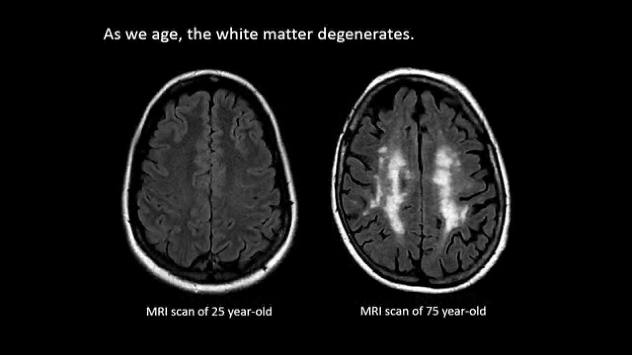
Ct scans and mri scans can show the loss of brain mass associated. Brain with dementia healthy older brains also work diff erently than y ounger brains. The most common types of brain scan you might encounter are magnetic resonance imaging (mri) and computed tomographic (ct) scans. Mri scans of 27 patients with probable alzheimer's disease (mean age 68.2 years), 31 patients with vascular dementia (mean age 69.9 years) and 18 normal controls (mean age 66.3 years) were compared to evaluate possible distinguishing parenchymal abnormalities among these groups.
Doctors will find it easier to tell whether a patient has alzheimer’s disease or another kind of dementia with a new method of using mri scans, researchers from perelman.
An mri imaging slide presented by turner showed a stark contrast in brain size between normal and alzheimer's brains. A prospective study with pathological verification of diagnosis. As beate has said, the basic scan is to rule out other conditions, apart from alzheimer's disease which could be causing the dementia as it's not easy to diagnose alzheimer's disease from a scan sometimes, areas of the plaques can be seen in a detailed scan, such as a spect scan but these are not usually carried out as if nothing is seen on the ct or mri scan,. Also, cortical atrophy—degeneration of the brain's cortex (outer layer)—is common in many forms of dementia and may be visible on a brain scan.
Atrophy was quantitated by subjective rating, linear and volumetric measurements.
Alzheimer’s disease & dementia vs. Drawing on brain scans of 32 people with alzheimer’s disease, researchers concluded that tau might play a more direct role in brain damage. Doctors rely on ct and mri brain scans when examining patients with suspected dementia. Frontotemporal dementias are a group of afflictions that are identified by a significant reduction in the amount of matter in the frontal and temporal lobes of the brain.
In turn, they become larger than normal.
This kind of imaging serves as a. A new study shows that mri brain scans can help doctors tell which people with certain thinking and memory problems might go on to develop dementia with lewy bodies rather than alzheimer's disease. These structural changes within the brain are also aspects that brain scans can identify. Five to 10 percent of americans diagnosed with dementia could actually have a treatable condition known as normal pressure hydrocephalus or nph, according to experts in the field.
• a continuous shrinking of brain tissue.
However, higher levels of brain and cognitive reserve are well known to attenuate the strength of these correlations and highly intelligent ad patients can be clinically mild, but. Dementia or alzheimer's brain scan would show different patterns in an alzheimer's brain versus a normal brain. Scott turner, md, phd ; By lisa esposito , staff writer aug.
This is because of the changes in neuron function, due to the damage from the plaques and tangles that have been building up in the brain.
2, 2019 this ar ticle is based on r epor ting that features exper t sources including r. In her phd research hafkemeijer used mri scans to detect changes in brain networks that occur as a result of ageing and dementia.